chapter 13 Small Animal Forelimb
Lateral View
Dorsal to vertebral column.
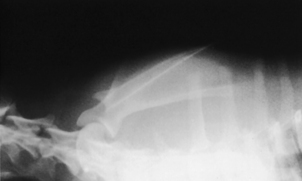
The best unobstructed view of the scapula is achieved by pushing the leg of interest dorsally so that the scapula is positioned dorsal to the vertebral column (Figs. 13-1 and 13-2).
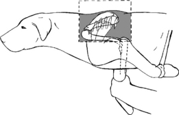
Figure 13-1 Correct positioning for the lateral view of the scapula positioned dorsal to the vertebral column.
The patient is placed in lateral recumbency with the affected limb closest to the cassette and held perpendicular to the spine. The limb is then pushed dorsally by grasping it firmly below the elbow and extending the elbow joint. With the elbow in extension, the joint cannot flex, allowing the scapula to be pushed dorsally. As the affected leg is pushed dorsally, the opposite leg is pulled ventrally. By pulling the opposite leg, the thorax becomes slightly rotated, which isolates the scapula dorsal to the body. At this point, the scapula should be seen bulging above the dorsal spinous processes of the thoracic vertebrae. Sedation is usually indicated for this view because of the firm manipulation necessary.
BEAM CENTER: Middle of scapula
Superimposed over cranial thorax.
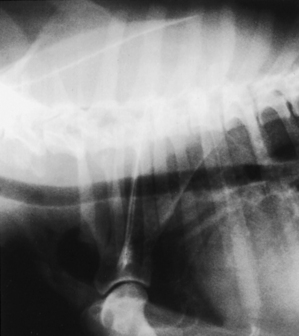
The view of the scapula superimposed over the cranial thorax is indicated for a patient that is in pain or when excessive manipulation may induce further injury (Figs. 13-3 and 13-4). The body of the scapula is placed over the radiolucent lung fields, allowing visualization of the majority of the bone. Although the entire scapula is not visible, this view is valuable for evaluation of the neck and body.
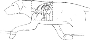
Figure 13-3 Correct positioning for the lateral view of the scapula superimposed over the cranial thorax.
Caudocranial View
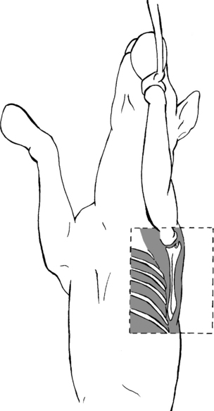
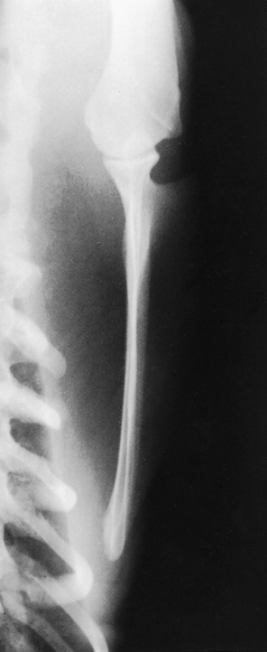
The patient is placed in dorsal recumbency (on its back) with both forelegs extended cranially (Figs. 13-5 and 13-6). The patient’s sternum should be rotated away from the scapula approximately 10 to 12 degrees, which alleviates any superimposition of the ribs of the thoracic cavity over the scapula and gives a clear, unobstructed view of the structure.
BEAM CENTER: Middle of scapula
MEASUREMENT: Cranioventral thorax where scapula is positioned
BEAM CENTER: Middle of scapula
Lateral View
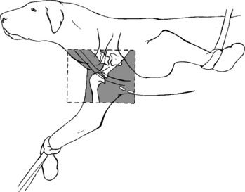
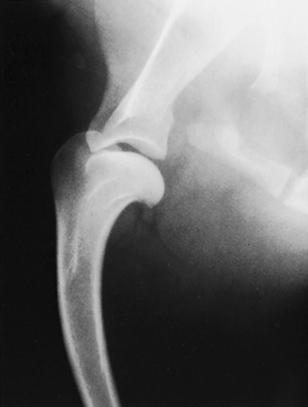
The patient is placed in lateral recumbency with the shoulder of interest closest to the cassette (Figs. 13-7 and 13-8). To alleviate any superimposition of structures over the shoulder, the leg must be extended cranially and ventrally to the sternum. The opposite limb is pulled in a caudodorsal direction, and the neck is extended dorsally. This gesture rotates the sternum slightly away from the shoulder joint. Care should be taken not to overrotate the thorax because the shoulder may be lifted off the cassette.
BEAM CENTER: To shoulder point
MEASUREMENT: Thickest area over shoulder joint
Caudocranial View
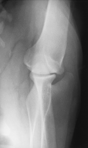
The position for the caudocranial shoulder is similar to the corresponding view of the scapula. The patient is placed in dorsal recumbency with both forelimbs extended cranially (Figs. 13-9 and 13-10). The limb should be extended so that the humerus is almost parallel to the cassette. Sedation may be necessary to allow such extension. As the forelimb is extended, care must be taken not to rotate the humerus. Any rotation would create an oblique view of the shoulder joint.
BEAM CENTER: To shoulder joint
Lateral View
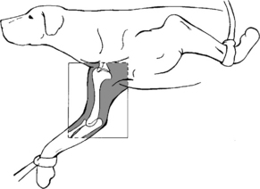
The patient is in lateral recumbency with the affected limb placed on the cassette. The leg is extended in a cranioventral direction, with the opposite limb drawn in a caudodorsal direction (Figs. 13-11 and 13-12). The head and neck should be extended dorsally. The field of view should include both the shoulder and the elbow joint with the humerus centered to the cassette.
BEAM CENTER: Center of humerus
