CHAPTER 51 Sinoscopy and Sinus Surgery in the Standing Horse
SINOSCOPY
Preparation
The paranasal sinuses can be explored endoscopically, with the horse standing, to determine the cause of signs of disease referable to the paranasal sinuses. The paranasal sinuses are examined with an arthroscope or sterile, flexible, video or fiberoptic endoscope. The endoscope is usually gas sterilized with ethylene oxide or disinfected in a glutaraldehyde-based solution. A tube of polyvinyl chloride pipe, one end of which has been capped, provides a convenient receptacle for the insertion tube of a flexible endoscope and the disinfectant. The accessory channel of the endoscope should be lavaged with disinfectant, and the insertion tube, including the accessory channel, should be rinsed with sterile water before the tube is inserted into the paranasal sinuses.
In preparation for endoscopy, the horse is sedated with detomidine (0.01 to 0.02 mg/kg, administered intravenously [IV], or 0.03 to 0.04 mg/kg, administered intramuscularly [IM]), and the sites at which the portals are to be created are prepared for surgery and desensitized by subcutaneous (SC) injection of local anesthetic solution. Endoscopic portals are made in the frontal or maxillary bone, through 2- to 3-cm longitudinal cutaneous incisions, by use of a 9.5-mm (3/8-in.) Galt trephine.∗ The incision is closed with staples or skin sutures after the endoscopic procedure.
Endoscopic Procedure
The conchofrontal sinus can be examined through a portal created in the frontal bone directly over the frontomaxillary aperture. The incision for the portal into the conchofrontal sinus is made in the skin overlying the frontal bone and is centered at a site two thirds of the distance from the dorsal midline to the medial canthus of the eye and 0.5 cm caudal to an imaginary line connecting both medial canthi. Structures that can be viewed directly through this portal include the frontal and dorsal conchal sinuses, ethmoid turbinates, frontomaxillary aperture immediately below the trephine hole, and nasomaxillary aperture at the rostral aspect of the frontomaxillary aperture. Other structures can be seen in the caudal maxillary sinus, including the infraorbital canal, apices of the 5th and 6th cheek teeth, maxillary septum, and entrance to the sphenopalatine sinus at the caudal aspect of the caudal maxillary sinus, medial to the infraorbital canal (Figure 51-1).

Figure 51-1 Sinoscopic view of the right conchofrontal and caudal maxillary sinuses of an 8-year-old horse with primary sinusitis. The bulla of the ventral conchal sinus (BVCS) is being broken down with a crocodile forceps inserted through the same trephine hole as the endoscope. CMS, caudal maxillary sinus; DCS, dorsal conchal sinus; IOC, intraorbital canal; NMAC, nasomaxillary aperture of the caudal maxillary sinus. See also Color Plate 2, following p. 256.
The maxillary septum prevents examination of the rostral maxillary and ventral conchal sinuses through a portal in the conchofrontal or caudal maxillary sinus, but the rostral maxillary sinus can be examined through a more rostral portal created in the maxillary bone. The incision for the portal into the rostral maxillary sinus is centered at a point 1 cm ventral to an imaginary line drawn between the infraorbital foramen and the medial canthus of the eye, midway between the rostral end of the facial crest and a point on the facial crest at the level of the medial canthus of the eye. The entrance to the ventral conchal sinus (the conchomaxillary aperture) and sometimes the rostromaxillary portion of the nasomaxillary aperture can be observed through this portal. The rostral maxillary sinus, a frequent site of infection caused by apical dental infection, is difficult to evaluate endoscopically because of its small size. The rostral maxillary sinus of young horses is especially difficult to evaluate because all or a portion of the long reserve crowns of the 3rd, 4th, and 5th cheek teeth occupy most of the sinus. Care should be taken when creating a portal into the rostral maxillary sinus of a young horse to avoid damaging the reserve crowns of the first and second maxillary molars, which reside completely or partially in this cavity.
The ventral conchal sinus, a frequent site of inspissated exudate, is difficult to evaluate endoscopically because it cannot be assessed directly through a trephine hole in the frontal or maxillary bone. The ventral conchal sinus, along with the rostral maxillary sinus, can be evaluated endoscopically through a portal into the conchofrontal sinus or caudal maxillary sinus after the cartilaginous bulla of the ventral conchal sinus is fenestrated with endoscopic guidance and an arthroscopic rongeur or crocodile-type forceps passed through the same portal as the endoscope (Figures 51-2 and 51-3). If fenestrating the bulla causes bleeding that obscures vision, sinoscopy should be repeated the next day. Fenestrating the bulla allows for more effective lavage of all compartments.
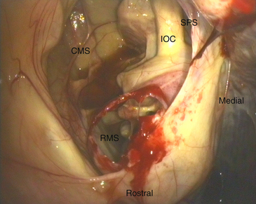
Figure 51-2 Sinoscopic view of the right conchofrontal and caudal maxillary sinuses of the same horse with primary sinusitis. The bulla of the ventral conchal sinus has been broken down, and the apex of the 5th cheek tooth can be seen in the rostral maxillary sinus. CMS, Caudal maxillary sinus; IOC, intraorbital canal; SPS, entrance to the sphenopalantine sinu; RMS, rostral maxillary sinus. See also Color Plate 3, following p. 256.
Stay updated, free articles. Join our Telegram channel

Full access? Get Clinical Tree


