Fig. 49.1
(a)–(c) Light micrographs of a xenograft tumor of Lu99 cells prepared with cryobiopsy show that there is little formation of ice crystal s (b, solid arrowheads) and flowing erythrocyte s are clearly observed in blood vessel s (b, solid arrow) near the tissue surface (a, solid arrows). Ice crystals can be found in the deeper areas (c, solid arrowheads). (d)–(k) Xenografted tumors of EBC-1 cells prepared with cryobiopsy (d, e), in vivo cryotechnique (IVCT; f, g), quick-freezing of fresh resected tissues (FQF; h, i), or immersion-fixation followed by dehydration (IMDH; j, k) show that open blood vessels (d, f, solid arrows) are clearly observed in regions near the tissue surface with cryobiopsy and IVCT (d, f, open arrows). Large pale tumor cell nuclei (e, g, solid arrowheads) and blood vessels (e, g, open arrowheads) with flowing erythrocytes (e, g, solid arrows) are also obvious. Blood vessel s are collapsed with FQF (h, solid arrows) near the frozen tissue surface (h, open arrow) and with IMDH (j, k, solid arrows). Large tumor cell nuclei are present with FQF (i, solid arrowheads), but the tumor cell nuclei are smaller and surrounded by damaged tissue with IMDH (k, solid arrowheads). Bars 50 μm (a, d, f, h, j) or 20 μm (b, c, e, g, i, k) (The figure is adapted from Ohno et al. [16])
Preparation artifacts are common in chemical fixation and dehydration methods, such as the formation of a chromatinic rim or nuclear shrinkage [18]. The artifacts, including cellular and nuclear aggregation and shrinkage , are prevented using freeze-substitution [19, 20]. The diagnosis and evaluation of prognosis is often associated with the histological features of tumor tissues, such as nuclear size, nucleocytoplasmic ratio, pleomorphism, and cytoplasmic anisocytosis [21–23]. It is possible that the cryotechniques can preserve the nuclear, cellular, and tissue structures of living tumors and contribute to the clinical diagnosis with fewer artifacts.
The modified Chalkley method was used to quantitatively measure the von Willebrand factor-positive blood vessel volume on paraffin sections [16]. The blood vessel volume in specimens prepared with IVCT or cryobiopsy was significantly larger compared with FQF or IMDH. The significant decrease in the blood vessel volumes was attributable to ischemia of tumor tissues which was inevitable upon tissue resection in FQF and IMDH. Evaluation of blood vessels can result in better estimates of clinical outcomes [24, 25]. The technical artifact s of conventional tissue preparation methods may influence the blood vessel volume or density. Cryobiopsy and IVCT will support the histopathological and ultrastructural examination of blood vessels in tumors without those artifacts .
49.3 Visualization of IgM -Immunopositive Blood Vessels
With cryobiopsy and IVCT, the immunostaining for IgM in tumor tissues was restricted in blood vessel s with flowing erythrocyte s [16]. Abundant von Willebrand factor (vWF)-immunopositive blood vessels were also observed in the same areas of serial sections. The IgM immunoreactivity was detected in some of the vWF-immunopositive blood vessels. In cryosections of the xenograft tumors, the intravenously injected BSA was clearly observed in blood vessels of the tumor tissues and capillaries of the capsular connective tissues around the tumor periphery (Fig. 49.2) [16]. The BSA was localized in some of the blood vessels immunopositive for vWF and collagen type IV in the vascular basement membrane s (Fig. 49.2). The immunoreactivity of BSA and IgM was detected in the same blood vessels of the tumor masses (Fig. 49.2). Therefore, the functional blood vessels with blood circulation can be visualized with IgM immunoreactivity in specimens prepared with both IVCT and cryobiopsy in the tumor tissues.
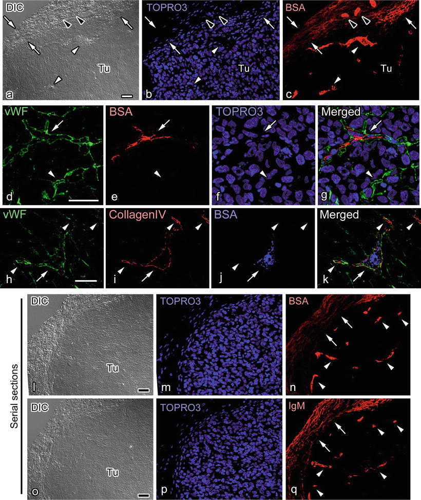
Fig. 49.2
(a)–(c) In the cryosections of a Lu99 cell xenograft tumor prepared using in vivo cryotechnique , bovine serum albumin (BSA, red) is immunolocalized in blood vessel s (solid arrowheads) around the tumor masses (Tu) and occasionally leaked in the adjacent connective tissues (open arrows). BSA immunoreactivity is also observed in blood vessels entering the tumor (open arrowheads). (d)–(g) BSA (red) is immunolocalized in some blood vessels (open arrows) positive for von Willebrand factor (vWF, green) but not in the other vWF-positive blood vessels (open arrowheads). (h)–(k) BSA immunoreactivity (blue) in some blood vessels (open arrows) labeled with vWF (green) and collagen type IV α2 chain (red) but not in others (open arrowheads). (l)–(q) Immunostaining for BSA (l–n) or IgM (o–q) in serial sections shows that BSA (n) and IgM (q) immunostaining appears similar in connective tissues around the tumor mass (n, q, open arrows) and blood vessels in the tumors (n, q open arrowheads). Cellular nuclei are labeled with TOPRO3 (b, f, m, p, blue). Bars 30 μm (The figure is adapted from Ohno et al. [16])
The relationship between fewer IgM -immunopositive blood vessel s and hypoxic states in the tumor cells was examined with IVCT [16]. The distribution of the IgM-immunopositive blood vessels and VEGF expression, a signal factor induced by hypoxic stimuli, was compared [26]. Serial sections of tumor tissues stained with hematoxylin-eosin or immunostained for IgM, vWF, and VEGF showed that the distribution of IgM immunoreactivity and erythrocytes was similar and could represent blood circulation (Fig. 49.3). The immunostaining for vWF was observed throughout the tumors (Fig. 49.3). On the other hand, the distribution of VEGF showed a clear contrast to that for IgM (Fig. 49.3).
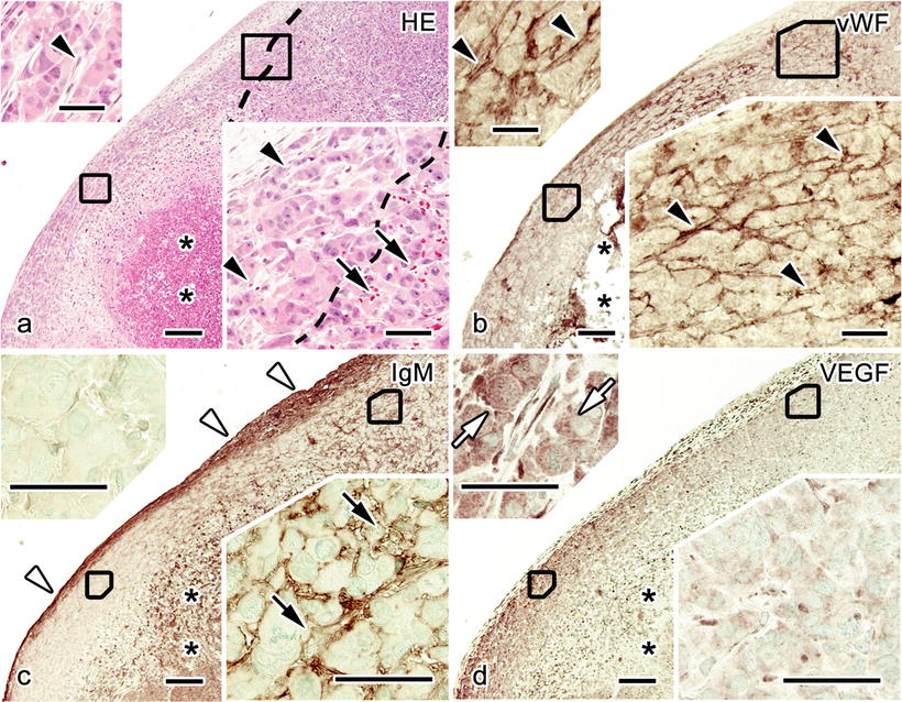
Fig. 49.3


(a)–(d) Light micrographs of serial paraffin sections obtained from an EBC-1 cell tumor and stained with hematoxylin-eosin (HE; a) or immunostained for von Willebrand factor (vWF; b), immunoglobulin M (IgM ; c), or vascular endothelial growth factor (VEGF; d). Erythrocytes (a, solid arrows) and IgM immunoreactivity (c, solid arrows) are observed in restricted areas, whereas vWF is immunolocalized throughout the tumor (a, b, solid arrowheads). VEGF immunoreactivity is stronger in the regions with little IgM immunostaining (d, open arrows). Open arrowheads: IgM-immunopositive connective tissues. Asterisks: necrotic areas. Bars 200 μm or 50 μm in insets (The figure is modified from Ohno et al. [16])
Stay updated, free articles. Join our Telegram channel
Full access? Get Clinical Tree
