14 Rodent, Lagomorph, and Ferret Dentistry
When you have completed this chapter, you will be able to:
• Define the term heterodont dentition and explain how the term applies to ferrets, rodents, and lagomorphs.
• Define the terms aradicular hypsodont, brachyodont, elodont, and peg teeth and explain how the terms apply to ferrets, rodents, and lagomorphs.
• Describe the unique structure of the peg teeth in lagomorphs and hypsodont cheek teeth of rodents and lagomorphs.
• List the dental formulas of the hamster, rat, gerbil, guinea pig, ferret, and adult rabbit.
• List the equipment needed for dental care of rodents, lagomorphs, and ferrets.
• List and describe common oral problems and diseases in rodents and lagomorphs.
• List and describe common oral problems and diseases and treatments in ferrets.
Dental Anatomy
Dentition in the Rodent, Lagomorph, and Ferret
Type of teeth in the ferret
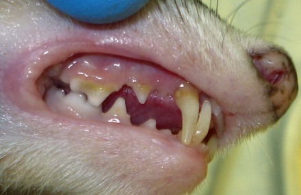
Ferrets have long, thin canine teeth that form a tight dental interlock when the mouth is closed. Incisor teeth are small with three incisor teeth in each quadrant. The maxillary fourth premolar and mandibular first molar teeth are sectorial, closing in a scissor-like fashion (Figure 14-1). Hourglass-shaped maxillary first molar teeth are a common dental finding of the family Mustelidae. The relatively small size of the teeth and oral cavity, coupled with the animal’s active nature, makes it difficult to thoroughly assess oral health in conscious ferrets, and therefore oral pathology can easily be overlooked in the conscious patient. Ferrets, being carnivores, have a highly specialized brachyodont dentition.
Adult Rodent Dental Formulas
Dental formulas of common rodents are as follows:
Hamster: 2 × (I 1/1, C 0/0, P 0/0, M 2-3/2-3) = 12 to 16 total teeth
Rat: 2 × (I 1/1, C 0/0, P 0/0, M 2-3/2-3) = 12 to 16 total teeth
Gerbil: 2 × (I 1/1, C 0/0, P 0/0, M 3/3) = 16 total teeth
Guinea pig and chinchilla: 2 × (I 1/1, C 0/0, P 1/1, M 3/3) = 20 total teeth
Squirrel: 2 × (I 1/1, C 0/0, P 1-2/1, M 3/3) = 20 to 22 total teeth
Adult Lagomorph Dental Formulas
The adult lagomorph dental formulas are as follows:
Rabbit: 2 × (I 2/1, C 0/0, P 3/2, M 2-3/3) = 26 to 28 total teeth
Instruments and Equipment Used to Treat Rodents, Lagomorphs, and Ferrets
Towels are helpful for restraint and for maintaining normothermia during procedures.
Anesthesia is available in two forms: injectable and inhalant. Injectable medications such as ketamine and acepromazine usually result in a longer recovery time than inhalants. Some injectables are reversible. Inhalants such as isoflurane and sevoflurane are preferred for maintenance of anesthesia for most procedures. Anesthesia masks can be used for induction of anesthesia prior to intubation or for the maintenance of anesthesia using an off-and-on mask approach during the actual dental procedure. Induction may also be administered in an anesthesia chamber, though small mammals may hold their breath due to the odor of the inhalant. Anesthesia and oxygen may also be delivered through an endotracheal tube. A laryngoscope with a small pediatric straight blade is used for endotracheal intubation to maintain the airway and administer anesthesia. Endotracheal tubes in sizes 1.5, 2.0, 2.5, or 3.0 Cole or a standard endotracheal tube may be used depending on the patient’s size. A wire stylet placed in the tubes makes passage much easier (Figure 14-2).
An otoscope with ear cones or a nasal speculum may be used to visualize the teeth and oral cavity of small mammals. In addition, these can be used as an aid in passing endotracheal tubes.
Intravenous (IV) catheters (12 to 14 gauge) can be used as an endotracheal tube for smaller rodents if the stylet is removed. These catheters can often be passed with the use of an otoscope as a laryngoscope.
Umbilical tape is used to anchor the endotracheal tube by snugly tying it to the tube and then behind the pet’s head.
Cotton-tipped applicators are used during dental procedures to clear the oral cavity of saliva, moisture, and debris.
Explorers/probes are used to examine teeth and their attachment structures.
Handpieces, both high-speed and low-speed, can be used with burs to shape, trim, prepare, or extract teeth.
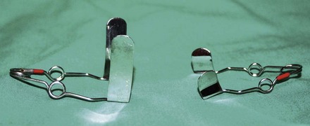
Cheek retractors are single-bladed instruments that retract the cheek on a single side or double-bladed instruments with a spring wire that spread both buccal folds at the same time (Figure 14-3).
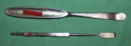
Tongue retractors are generally instruments with a single, flat blade used to move the tongue away from the area to be inspected or treated (Figure 14-4).
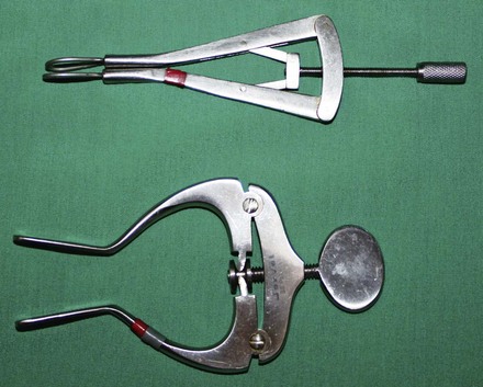
Mouth gags aid in keeping the mouth open during inspection and treatment. Most are spring activated or they may expand with a screw device (Figure 14-5).
Burs are used for removal of hard tissue. The most common types are diamond burs, carbide round burs, carbide tapered fissure burs, and white stone points. A cylindrical diamond bur works well for cheek teeth and incisor teeth. Carbide burs are more aggressive than diamond burs and white stones. Of the carbide burs, the 701L tapered fissure and #1 and #2 round ball burs are helpful. Of the white stone abrasive points, the flame-shaped point works well. Friction grip (FG) burs may be used on high-speed handpieces when working in the rostral oral cavity, but straight (HP) burs are necessary for use on the cheek teeth of most rabbits and rodents due to poor accessibility with the high-speed handpiece. A bur guard can be used to protect soft tissues from being traumatized by the bur.
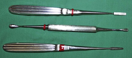
Files/rasps (sometimes referred to as “floats”) are instruments used to level an abnormally uneven occlusive table of the teeth. The hand floats used in the small mouths of rodents and lagomorphs are typically either modified bone files or rasps. The rasps cut both on the push and pull stroke, whereas the files typically cut only on the pull stroke. The BF4 bone rasp (Cislak, Inc., Glenview, IL) and the Howard 12 bone file (Butler-Schein, Inc., Port Washington, NY) are two instruments that can be used for floating cheek teeth (Figure 14-6). The rabbit molar rasp is a float specifically developed for use in rabbits. Most have a large handle for better control and a working end that is similar to a bone rasp. The J-41r (Jorgensen Labs, Inc., Loveland, CO) is a smaller diamond-coated rasp that works well for final smoothing of jagged tooth surfaces after use of a bur or larger file to reduce an overgrown tooth.
Molar or cheek teeth cutters may be modified hard-tissue nippers, pin and wire cutting pliers, or side-cutting rongeurs. However, some have been developed specifically for use in rabbits (Figure 14-7).
Elevators are used to work circumferentially around a tooth in the PDL space to aid in the tooth’s removal. Blunted injection needles, sizes 18- to 25-gauge, can often be used for this function in rodents and lagomorphs. However, standard elevators can also be used, such as No. 1 and 2 winged elevators, 301 apical elevators, and Crossley Rabbit Luxators (Jorgensen Labs, Inc., Loveland, CO).
Extraction forceps are used to grasp teeth loosened by elevation or severe disease. Most incisors can be handled with small animal extraction forceps. For the cheek teeth a small 90-degree angled Halstead mosquito forceps or an angled root tip forceps can be useful. Some extraction forceps are specifically designed for use in rabbits.
Bone substitutes are placed into areas in which periodontal or endodontic disease has resulted in bone loss. These grafts may be combined with an antibiotic or other medicament when placed in a bony void.
Antibiotic-impregnated beads may be placed for treatment of refractory infections. They may be nonabsorbable such as polymethylmethacrylate beads. Calcium phosphate antibiotic impregnated beads are typically absorbed within weeks after placement.
Paper points are commonly used to apply medicaments to exposed pulps.
Calcium hydroxide is commonly used in either a powder or paste form. It is placed over the exposed pulp of a tooth in an attempt to maintain its vitality. Its use has also been described as a packing material in infected draining abscesses, though this use can cause severe soft tissue necrosis due to its high pH.
Restoratives used in small mammal and exotic pets are ordinarily a temporary filling material such as Cavit (3M ESPE, St. Paul, MN), reinforced zinc oxide–eugenol (ZOE) cements, or glass ionomer restorative materials.
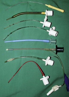
FIGURE 14-2 Standard, Cole-type, and handmade endotracheal tubes and a stylet for use during placement.
Stay updated, free articles. Join our Telegram channel

Full access? Get Clinical Tree


