Chapter 11 Retina, Choroid, Sclera
INTRODUCTION
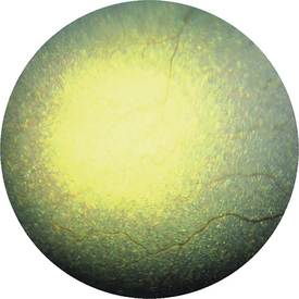
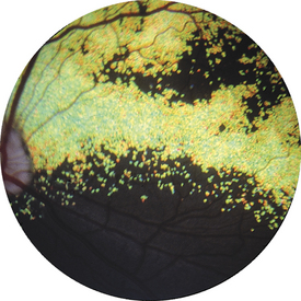
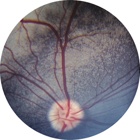
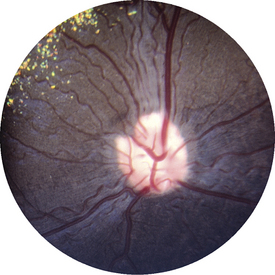
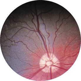
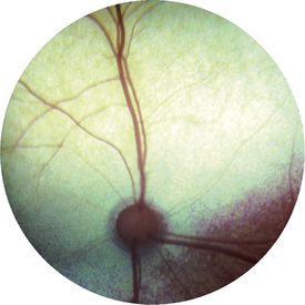
Figure 11-97 Rod-cone dysplasia type 1 in a 14-month-old Irish setter—the same dog as shown in Figures 11-98 and 11-99. This photograph shows the peripheral tapetum and marked tapetal hyperreflectivity and vessel attenuation.
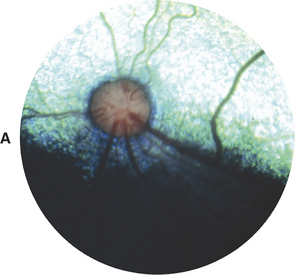
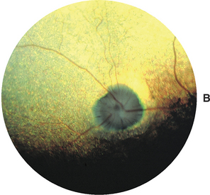
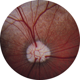
Figure 11-98 A-B A, Rod-cone dysplasia type 1 in an Irish setter at 4 months of age. Note the juvenile fundus, which ophthalmoscopically appears normal. B, Rod-cone dysplasia type 1 in the same dog at 10 months of age. This photograph is of the same dog as shown in Figures 11-97 and 11-99. Tapetal hyperreflectivity, vessel thinning, and optic atrophy can be seen.
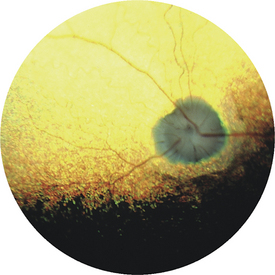
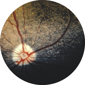
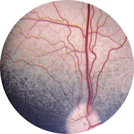
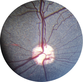
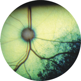
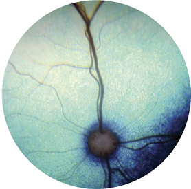
Figure 11-99 Rod-cone dysplasia type 1 in a 14-month-old Irish setter—the same dog as shown in Figures 11-97 and 11-98. Tapetal hyperreflectivity, vessel thinning, optic atrophy, and glial proliferations at edge of the nerve (gliosis) can be seen.
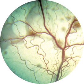
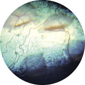
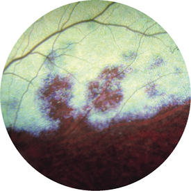
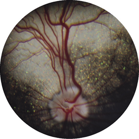
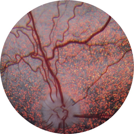
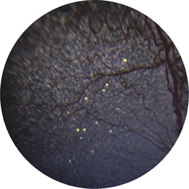
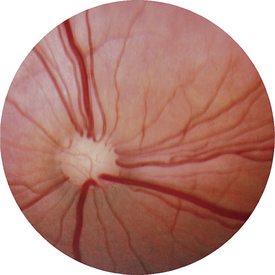
Figure 11-185 Mastiff (2 years old) with retinal dysplasia, with bullous retinal detachments and ventral gravitation of red-colored material. Same dog as shown in Figures 11-184 and 11-186.
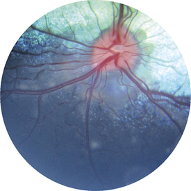
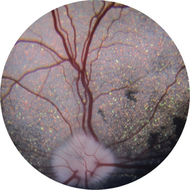
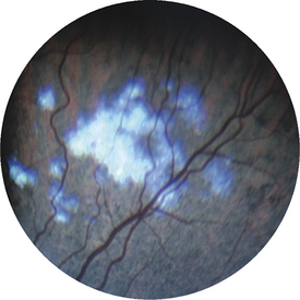
Figure 11-186 Mastiff (2 years old) with retinal dysplasia in the nontapetum. Same dog as shown in Figures 11-184 and 11-185.
NORMAL FUNDUS IN DOGS
Normal Tapetum in Dogs
When a dog’s tapetal fundus is viewed ophthalmoscopically, retinal vessels are superimposed on the highly colored tapetum. The only visible components of the retina are the blood vessels. The RPE in this region is nonpigmented to allow light to reach and be reflected by the tapetum. Therefore, the RPE layer is not visible either. The tapetum is visible as the aggregate of numerous tapetal cells that overlap one another to make a layer of variable thickness, depending on the location in the posterior segment, and varies between eyes according to the amount of pigment in the eye because the tapetum consists of modified pigment cells. The tapetum may be gray, violet, blue, green, yellow, or reddish orange. The surface area of the posterior segment covered by the tapetum also may vary, tending to occupy a larger area in larger dog breeds (and including the optic nerve in the tapetal area) and a smaller area (usually more on the temporal area of the fundus) in small breeds. Some dogs have no tapetum at all. The border of the tapetal and nontapetal fundus may be sharply defined (usually more likely in short-coated breeds) or irregular with interlocking of tapetal and nontapetal regions (in longer-coated animals). Dogs of same breeds often have the same tapetal appearance and often color.
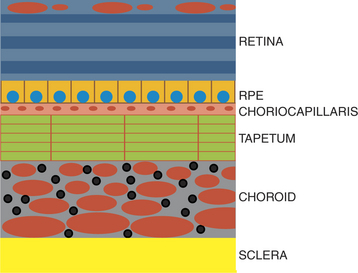
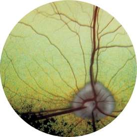
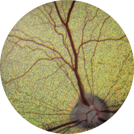
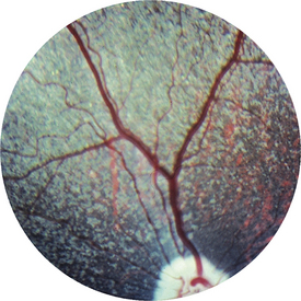
Figure 11-3 Normal retina with a speckled tapetum, the result of variable thickness of the tapetal cell layer.
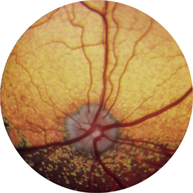
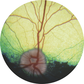
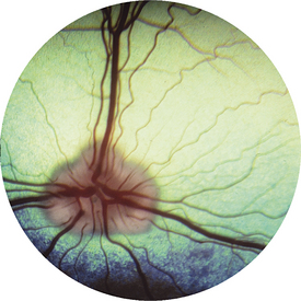
Figure 11-7 Normal fundus in a German shorthaired pointer with a large amount of optic nerve myelination.
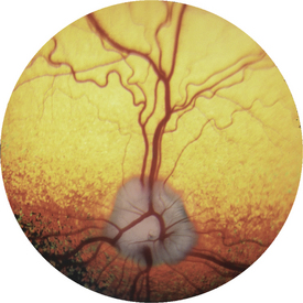
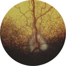
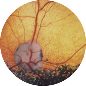
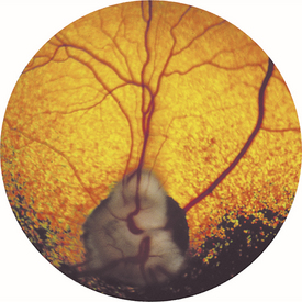
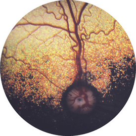
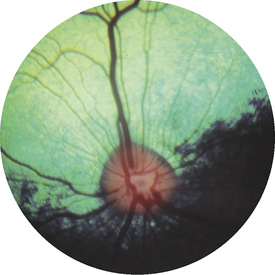
Figure 11-13 Normal retina. Extension of myelin out from the disc is limited, and a small pigmented conus surrounds the disc.
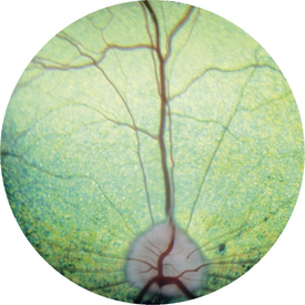
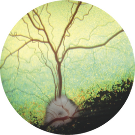
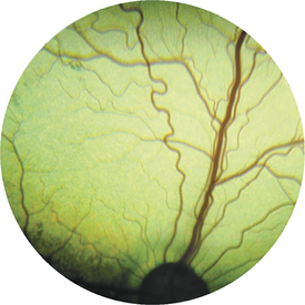
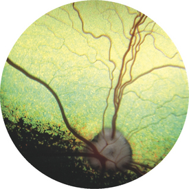
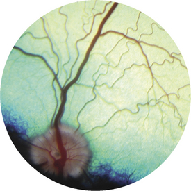
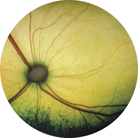
Figure 11-18 Normal retina. The vessels over the optic nerve are prominent; the arterioles are tortuous but normal.
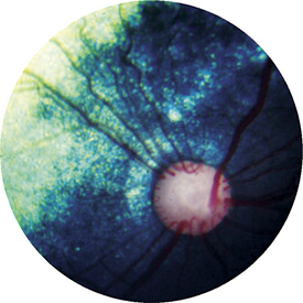
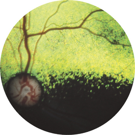
Figure 11-20 Normal retina. Pigment varies at the tapetal-nontapetal junction; pigment is present in the tapetum.
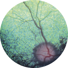
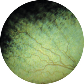
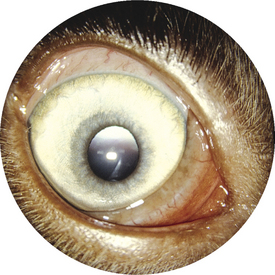
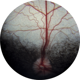
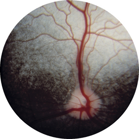
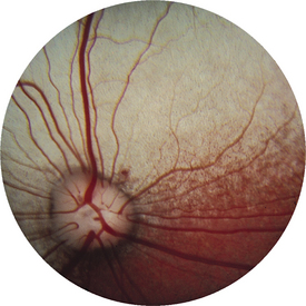
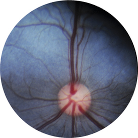
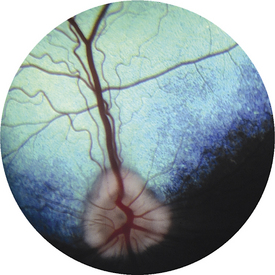
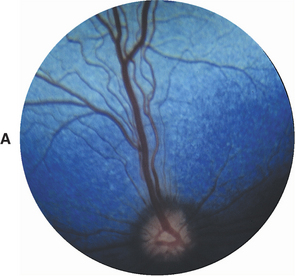
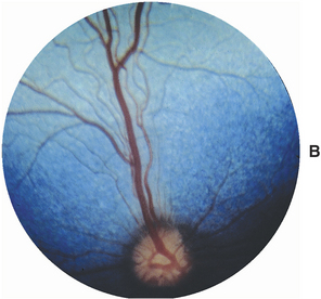
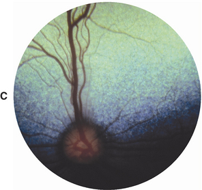
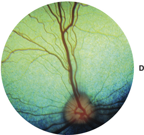
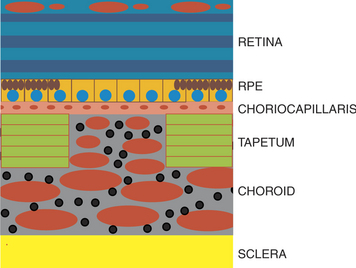
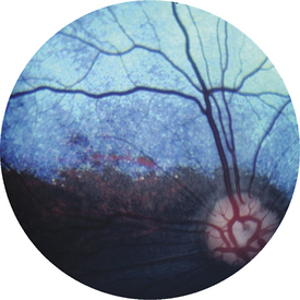
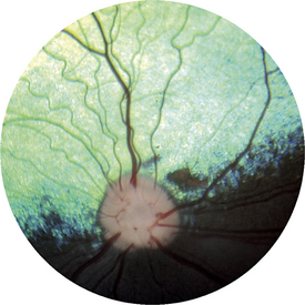
Figure 11-32 Normal retina. The tapetum has thinned areas through which choroidal vessels are showing.

Normal Nontapetum in Dogs
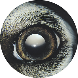
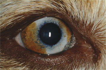
Figure 11-50 Normal dark-colored canine iris, which would be associated with a well-pigmented fundus.
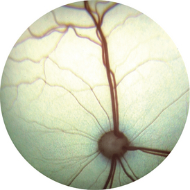
(Courtesy Dr. Robert Playter.)
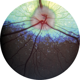
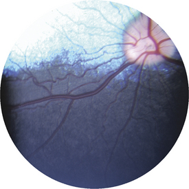
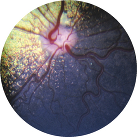
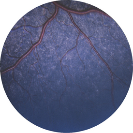
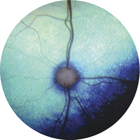
Figure 11-55 Normal dog nontapetum; the blue color is the effect of the camera flash distributed across the retina. This view is of a more peripheral area of the nontapetum seen in Figure 11-53.
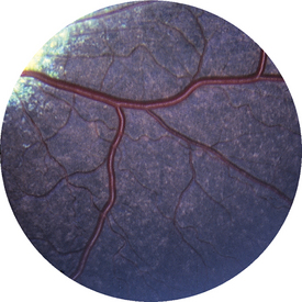
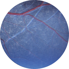
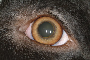
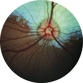
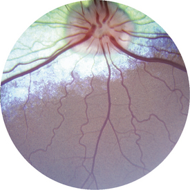
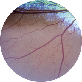
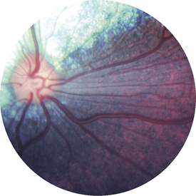
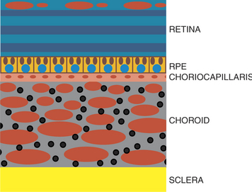
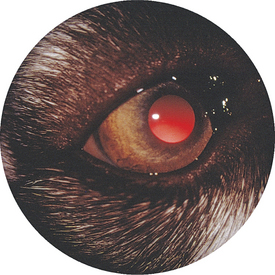
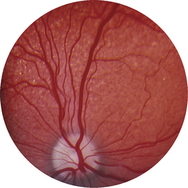
Figure 11-64 Normal Australian shepherd. Development of the tapetum is very limited, most evident in the temporal fundus.
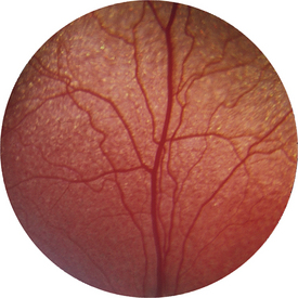
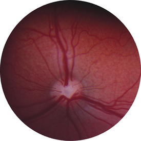
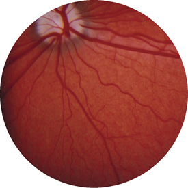
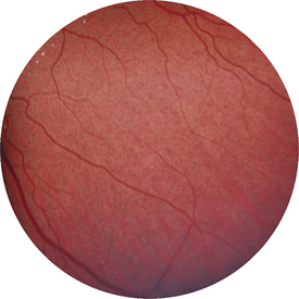
Figure 11-68 Nontapetum in a normal Australian shepherd. Distinct choroidal vessels can be seen inferiorly in the far periphery.
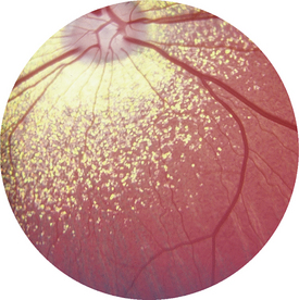
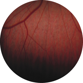
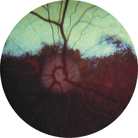
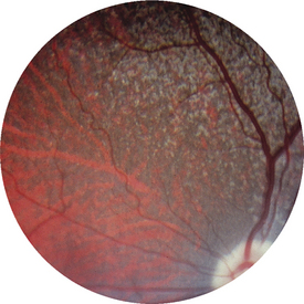
Figure 11-73 Normal merle collie. Choroidal vessels are visible on left side of photograph.
(Courtesy Dr. E. Dan Wolf.)
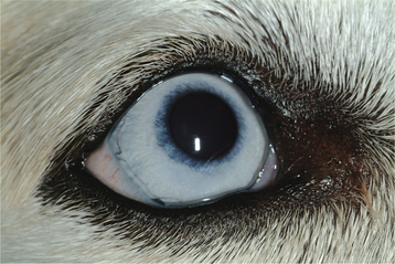
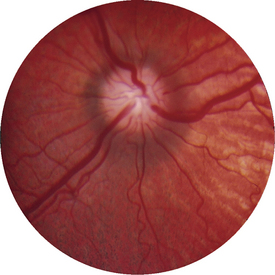
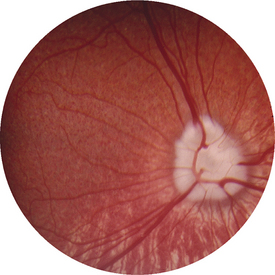
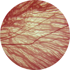
Figure 11-79 Normal dog. Choroidal vessels are visible throughout the fundus. More peripheral area of the same fundus as shown in Figure 11-78.
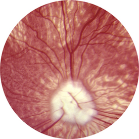
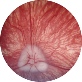
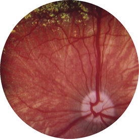
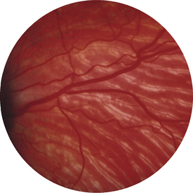
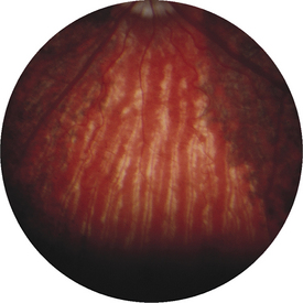
Figure 11-84 Australian shepherd from Figure 11-83. Some areas of RPE are normally pigmented; other areas are nonpigmented, and choroidal vessels can be seen.
NORMAL FUNDUS IN CATS
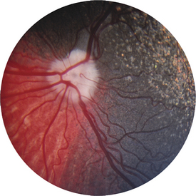
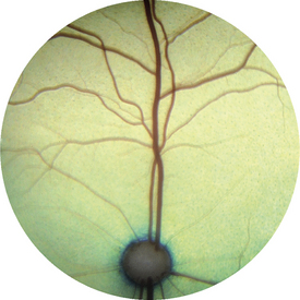
Figure 11-87 Normal cat. The tapetal color is typical, and the optic nerve is surrounded by tapetum.
< div class='tao-gold-member'>
Stay updated, free articles. Join our Telegram channel

Full access? Get Clinical Tree


