Chapter 6 Respiratory Diseases
Canine Distemper Virus
The virus is most commonly transmitted by aerosol exposure.2 Direct contact with conjunctival and nasal exudates, urine, feces, and skin can also cause infection.21 Ferrets shed virus in all body excretions, and shedding begins about 7 days after exposure.2 Fomites are also implicated in transmission; on gloves, the virus is viable for up to 20 minutes.21 Once in a ferret’s body, the virus appears to spread by viremia.43 The incubation period in ferrets is typically 7 to 10 days, although incubation periods of up to 56 days have been reported in natural infections.21,44
History and Physical Examination
Infection with CDV should be suspected in any unvaccinated, exposed ferret showing compatible clinical signs. Unvaccinated ferrets of any age are equally susceptible to this disease. In dogs, pyrexia develops 3 to 6 days after infection with CDV and is soon followed by anorexia and a serous nasal discharge.2 A serous ocular discharge then appears; this discharge quickly becomes mucopurulent.
In ferrets, the first sign of disease is usually a papular dermatitis on the chin, followed by a cheilitis characterized by swelling and crusting. These changes may be accompanied by dermatitis on the anus and inguinal area,21 which is orange-tinged in some ferrets. Other clinical signs are anorexia, depression, dyspnea, pyrexia, photophobia, pruritus, blepharospasm, and abundant mucopurulent oculonasal discharge. Hyperkeratosis of the planum nasale and footpads (Fig. 6-1) often occurs. Vomiting and diarrhea, which are seen in dogs with CDV, are uncommon in ferrets.
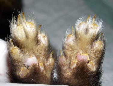
Fig. 6-1 Hyperkeratotic footpads in a ferret diagnosed with canine distemper virus (CDV).
Photograph courtesy of Dr. David Perpinan.
The respiratory system is the preferred site for the virus to replicate.43 Secondary bacterial infections, which are responsible for many of the severe respiratory symptoms and death, are caused by the immunosuppressive effects of the virus.57
Seizures and blindness are common in dogs with CDV infection,57 and neurologic signs may manifest without previous systemic signs.2 In ferrets with advanced CDV infection, incoordination, torticollis, and nystagmus can be present.2
Diagnosis
In the past, most laboratory tests available for the diagnosis of distemper were hampered by low sensitivity, low specificity, or both. A plasma sample can be submitted to measure an antibody titer against CDV. However, because both infected and vaccinated ferrets can have a positive titer, a positive result is not diagnostic of disease. In practical terms, if a ferret that has not been vaccinated has a positive titer, this test can confirm CDV infection. A fluorescent antibody test can be done on conjunctival smears, mucous membrane scrapings, or blood smears to identify CDV antigen in cells.21 However, this test is useful only in the first few days of disease, and false-negative results are possible. Modified live viral strains used for vaccination do not interfere with this test.57
Polymerase chain reaction (PCR) assays have been developed and may be used for ante- or postmortem diagnosis. Recent research suggests that nested PCR assays are more sensitive for antemortem diagnosis than reverse transcriptase PCR (RT-PCR).30,51 Samples of blood, urine, feces, or tissues or deep pharyngeal swabs should be submitted to commercial laboratories for CDV PCR testing. False-positive results may be seen in the first few weeks after vaccination with modified live vaccines. Killed and vector-recombinant vaccines do not interfere with PCR testing.
A positive postmortem diagnosis can be made by fluorescent antibody staining of imprints from lymph nodes, bladder epithelium, and cerebellum.2 Histopathologic examination of affected cells can also confirm the disease. Inclusion bodies of CDV are usually intracytoplasmic but can be intranuclear. Inclusions are generally found in the epithelial cells of the trachea, urinary bladder, skin, gastrointestinal tract, lymph nodes, spleen, and salivary glands.21 Diffuse interstitial pneumonia may be present. In the central nervous system, inflammatory cell invasion with demyelination is observed.2
History of exposure and clinical signs can be highly suggestive of infection. Nonspecific test results can include leukopenia, alpha- and beta-hyperglobulinemia, and radiographic evidence of lung congestion or consolidation.21,44,57 Nonregenerative anemia and increased serum levels of alpha and beta globulins are the most common routine laboratory changes.44
Treatment
No specific treatment exists for CDV infection in ferrets, and the mortality rate may be up to 100%. Death generally occurs 12 to 16 days after exposure to ferret-adapted CDV strains and 21 to 35 days after exposure to canine wild virus strains. Euthanasia of affected ferrets is usually the most humane option. Palliative treatment consists of supportive care and antibiotics for secondary bacterial infections. Administration of anti-canine distemper hyperimmune serum may be useful if given early in the course of the disease.44
Prevention
Vaccination is the best way to prevent CDV infection in ferrets. However, CDV vaccines are insufficient to induce protection in very young ferrets because of interference from maternal antibodies acquired via colostrum in the first few days of life. Because CDV is closely related to MV, vaccination with MV vaccine may offer cross protection at 5 to 6 weeks of age without interference from maternal antibodies.64 Otherwise, CDV vaccinations may be started at 6 or 8 weeks of age for kits from nonimmune or immune dams, respectively, and then continued every 3 to 4 weeks until the kits are at least 12 to 14 weeks old. At present, the recommendation is to revaccinate yearly. However, results of currently ongoing research demonstrate that antibody titers may remain high for several years, suggesting that boosters could be given less frequently. In other species, titers of 1:32 are considered protective, and in a clinical trial involving 66 CDV-vaccinated ferrets, average antibody titers were found to be over 1:1000 a year postvaccination (HL Heller, 2009, personal communication).
Currently, only one CDV vaccine approved by the U.S. government is available for ferrets: PureVax Ferret Distemper Vaccine (Merial, Athens, GA). Production of Fervac-D (United Vaccines, Madison, WI), the only other approved ferret distemper vaccine, was permanently discontinued by the manufacturer. Avoid use of multivalent canine vaccines, which can be associated with adverse effects. Vaccine strains of CDV that have been propagated in cell lines of canine origin may induce distemper disease in ferrets. Signs of vaccine-induced distemper may include mild purulent upper respiratory tract infection with pyrexia that resolves in a week or progresses to fulminating distemper during the same time frame (see Chapter 2).
Anaphylactic reactions in ferrets have been reported after vaccination.24,40 Most of these reactions occur after vaccination with canine distemper vaccines. Most reactions usually happen within 30 minutes after vaccination, with clinical signs of vomiting, diarrhea, pale mucous membranes, weak pulses, tachycardia, and lethargy. If a reaction occurs, treat the ferret for anaphylactic shock with epinephrine, parenteral fluids, steroids, antihistamines, and oxygen therapy as needed. It is prudent to suggest that a ferret owner remain in the hospital for up to 30 minutes after CDV vaccination in case a reaction should occur. PureVax ferret distemper vaccine is a recombinant canarypox vector vaccine that appears to be less likely to cause anaphylaxis. A possible link between myofasciitis and distemper vaccination has been suggested.22
If an outbreak of CDV occurs in a group of susceptible ferrets, all affected animals should be removed and the healthy ferrets vaccinated immediately. However, vaccinating nonimmunized ferrets may not stop infection and subsequent death in the face of an outbreak.21
CDV is relatively labile and its infectivity is destroyed by heat, drying, detergents, and disinfectants.57 Routine cleaning and disinfection procedures effectively destroy CDV on hard surfaces.
Influenza
Ferrets are susceptible to infection from both influenza type A and B viruses of the class Orthomyxoviridae. Natural outbreaks or clinical cases of influenza in ferrets have occurred with common human influenza type A viruses, the human strain of pandemic H1N1 virus, and swine-origin H1N1 influenza virus.10,42,55,58 The pathogenicity of type B influenza virus in ferrets appears to be low. In a recent report documenting natural cases of pandemic H1N1 influenza in ferrets, transmission was most likely from infected humans in the household.58 The influenza virus H3N8 that emerged in dogs in 2004 is most closely related to the equine influenza virus, whereas influenza A viruses affecting ferrets appear to have a pattern of viral attachment more similar to avian and human influenza subtypes.15,62 Although there is a theoretical potential of the virus being transmitted from ferrets to humans,36 there is only one report, from the 1930s, that documents a probable transmission of virus to humans. In that report, an animal-passage influenza strain was inoculated into a laboratory ferret, and a laboratory investigator was infected after close contact with the animal.54 Ferrets have long been important animal models of transmission, pathogenicity, and treatment studies of influenza virus in humans.
Influenza virus is transmitted primarily by aerosol droplets from ferret to ferret or from human to ferret. The virus can be transmitted beginning at the height of pyrexia and continuing for the next 3 to 4 days.56 In ferrets as in people, influenza primarily causes upper respiratory disease. The different subtypes of influenza A viruses vary in virulence and likelihood of developing secondary bacterial infections, which accounts for the difference in severity of clinical signs.4,30,46
History and Physical Examination
Ferrets contract influenza after being exposed to infected people or other infected ferrets. All ferrets are susceptible to influenza, although the disease is typically more severe in neonates than in older ferrets. After a short incubation period, the body temperature increases and then decreases approximately 48 hours later.14,21,36,49 The fever may be biphasic throughout the course of the disease. Bouts of sneezing, epiphora, and a mucoid or mucopurulent nasal discharge are common. Clinical signs can appear within 48 hours of exposure.14,36 Affected ferrets may become lethargic and anorexic,56 and photophobia and conjunctivitis may be present.5 Neonates develop a much more severe upper respiratory tract infection than adults, and death may ensue from lower airway obstruction.9,53
Clinical signs involving the lower respiratory tract are less common than those of the upper respiratory tract. Infection of the lower respiratory tract is usually confined to the bronchial epithelium51 and results from secondary bacterial infection. Death can ensue from secondary pulmonary infection with Lancefield group C hemolytic streptococci.36 Neonates are more likely than older ferrets to develop bronchiolitis and pneumonia14 and to die from lower respiratory tract infection.51
Influenza virus can infect the cells of the intestinal mucosa and cause a limited enteritis.23 The potential for hepatic dysfunction has been described in ferrets infected experimentally with influenza.31 Hearing loss has also been associated with influenza infection in ferrets.48
Stay updated, free articles. Join our Telegram channel

Full access? Get Clinical Tree


