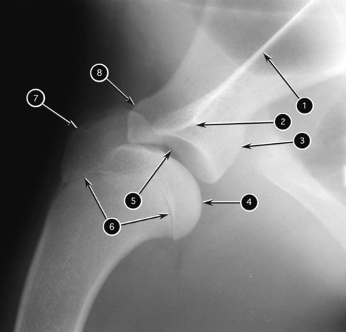
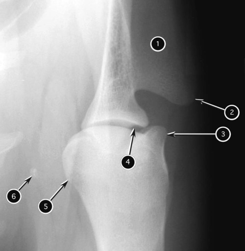
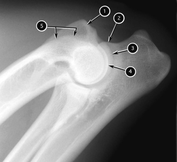
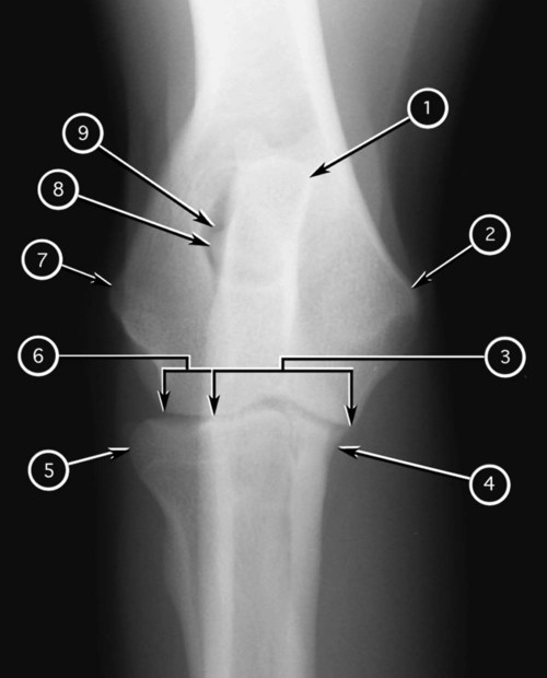
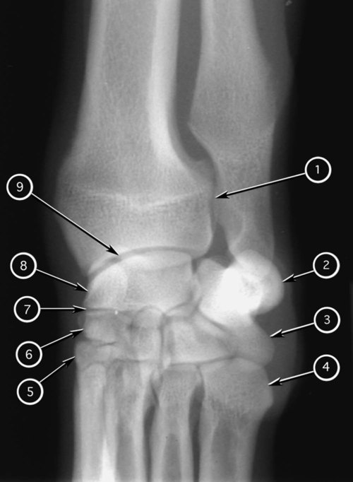
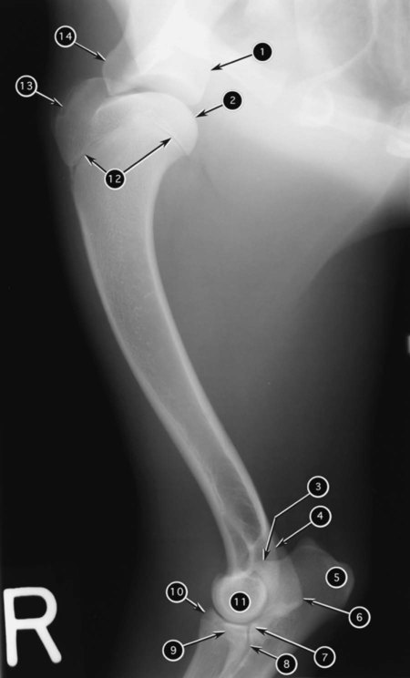
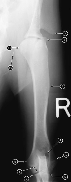
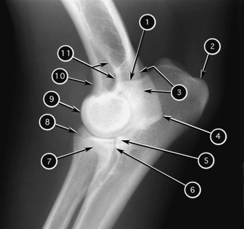
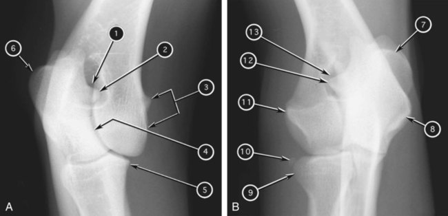
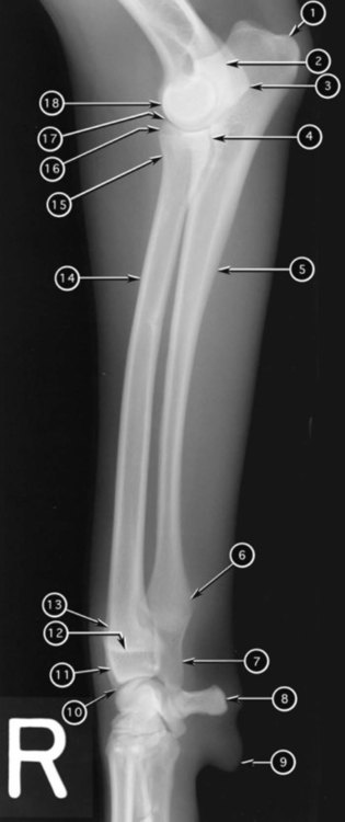
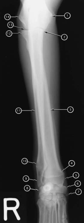
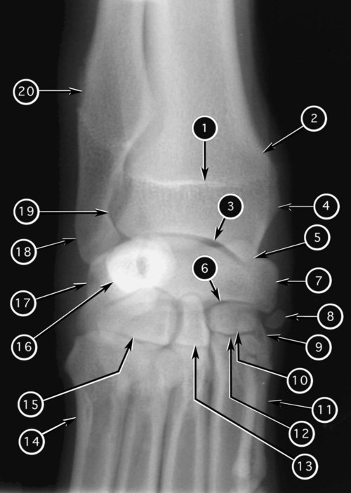
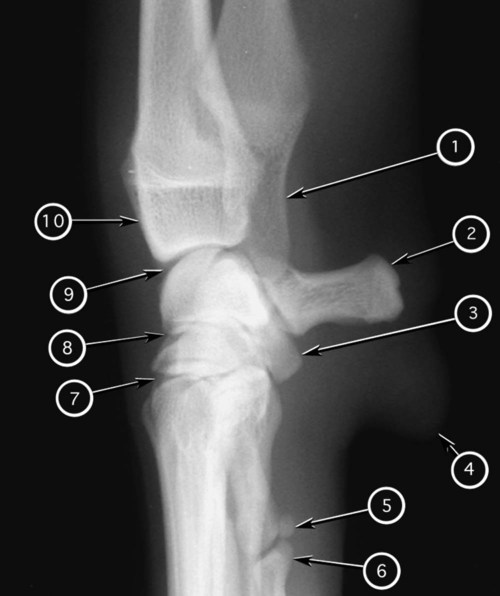
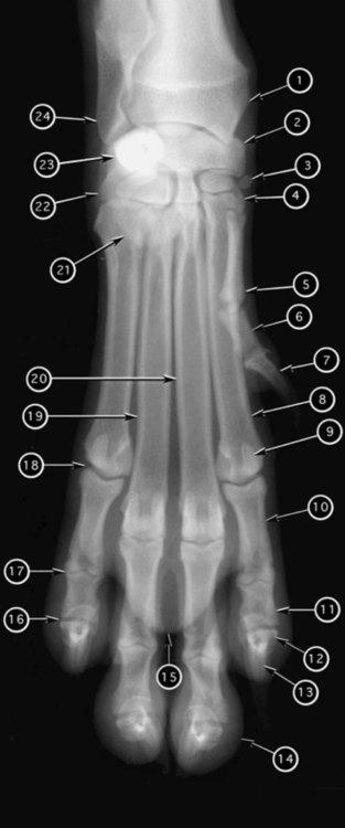
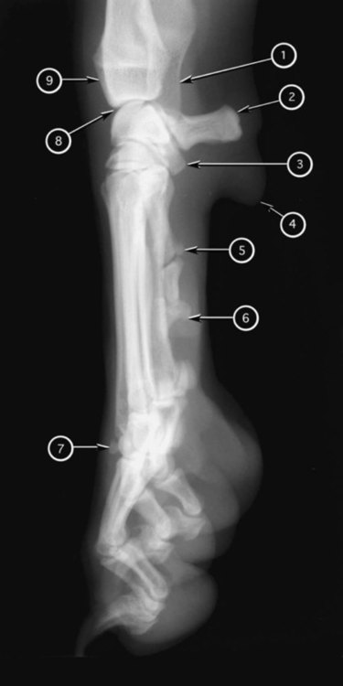
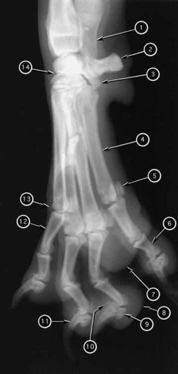
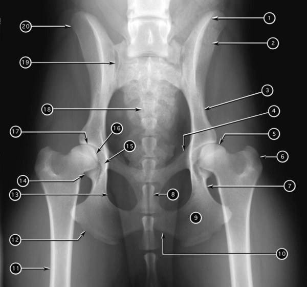
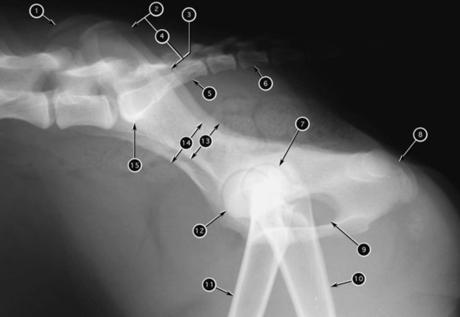
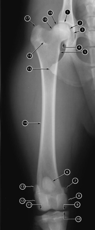
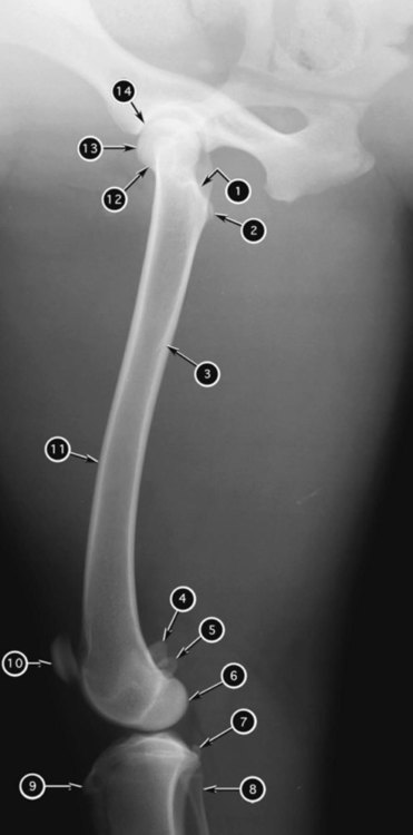
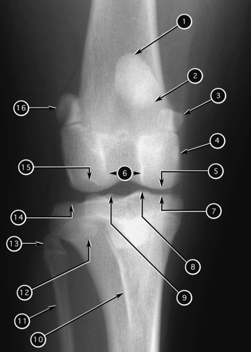
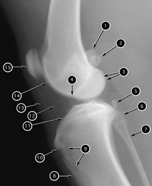
To use the roentgen sign method of recognizing abnormal radiographic findings effectively, an understanding of normal radiographic anatomy for the specific area of interest is necessary. The purpose of this chapter is to provide a limited reference for the radiographic anatomy of the appendicular skeleton. Refer to comprehensive textbooks on radiographic anatomy for more detailed information.1–3 The radiographic nomenclature used in this chapter was approved by the American College of Veterinary Radiology in 1983.4 Some equine images in this chapter (Figs. 13-30 through 13-49 and 13-54 through 13-57) have been taken from previous publications and are reproduced here with permission of the journals and author.5–7 2. Intermedioradial carpal bone 3. Sesamoid bone of abductor pollicis longus 6. Proximal phalanx of digit 1 9. Abaxial proximal sesamoid bone of digit 2 12. Unguicular crest of distal phalanx of digit 2 13. Unguicular process of distal phalanx of digit 2 16. Distal interphalangeal joint of digit 5 17. Proximal interphalangeal joint of digit 5 18. Metacarpophalangeal joint of digit 5 1. Distal epiphysis (styloid process) of ulna 5. Abaxial proximal sesamoid bone of digit 5 9. Flexor tubercle of distal phalanx of digit 4 10. Unguicular crest of distal phalanx of digit 4 11. Unguicular process of distal phalanx digit 3 12. Proximal phalanx of digit 2 13. Dorsal sesamoid bone of digit 2 1. Crest of right ilium (more magnified) 4. Left tuber sacrale (less magnified) 7. Left coxal joint (less magnified) 8. Superimposed right and left tubera ischiadica 9. Superimposed right and left obturator foramina 10. Body of right femur (more magnified) 11. Body of left femur (less magnified) 12. Head of right femur (more magnified) 13. Body of left ilium (less magnified) 14. Body of right ilium (more magnified) 1. Lateral sesamoid of gastrocnemius muscle 2. Medial sesamoid of gastrocnemius muscle 3. Medial and lateral condyles of femur 5. Sesamoid bone of popliteus muscle 9. Cartilage between tibial tuberosity and body of tibia
Radiographic Anatomy of the Appendicular Skeleton
![]()
Stay updated, free articles. Join our Telegram channel

Full access? Get Clinical Tree


Radiographic Anatomy of the Appendicular Skeleton