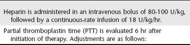
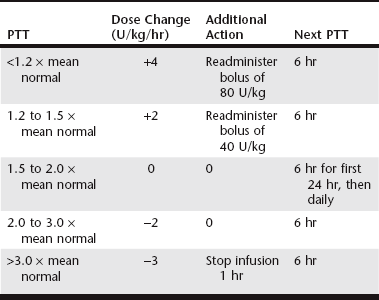
Pulmonary Thromboembolism

Chapter 166


Pulmonary Thromboembolism


Pulmonary thromboembolism (PTE) occurs when thrombi that develop in the venous circulation dislodge and become trapped in the pulmonary vasculature. If the degree of obstruction overwhelms compensatory mechanisms, respiratory (and sometimes hemodynamic) compromise ensues. Although the clinical manifestations of PTE may be mild, a seemingly high proportion of patients exhibit significant morbidity or even experience acute death. The prevention and management of PTE is the focus of this chapter. Critical to this therapy is a basic understanding of antithrombotic drugs.


PTE is a common complication of medical illness and surgery in humans, accounting for significant morbidity and over 100,000 deaths annually in the United States. Although less common in small animals, PTE is nevertheless an important clinical entity, and prevalence is highest in certain patient populations. For example, the mortality of canine immune-mediated hemolytic anemia (IMHA) is reportedly 50% to 70%, and PTE may account for up to 60% to 80% of these deaths.


PTE results from an increased thrombotic tendency, termed thrombophilia. Thrombophilia arises from some combination of three major mechanisms, as described by Virchow’s triad: endothelial injury, blood stasis, and prothrombotic alterations of the hemostatic system (hypercoagulability). The first feature is likely of greater importance in arterial thromboembolic pathophysiology and the latter in venous. Hypercoagulability results from platelet hyperaggregability, deficiencies of natural anticoagulants (antithrombin, protein C), or defective fibrinolysis.


The term hypercoagulable state refers to disorders in patients with underlying disease known to be associated with thrombophilia and an increased risk of thrombosis. In animals, these are acquired disorders, and the pathogenesis is generally multifactorial and complex. PTE in dogs is associated with IMHA, protein-losing nephropathy (PLN), neoplasia, necrotizing pancreatitis, hypercortisolism (hyperadrenocorticism and corticosteroid therapy), protein-losing enteropathy (PLE), cardiac disease (dirofilariasis, endocarditis, and cardiomyopathy), diabetes mellitus, sepsis, atherosclerosis, trauma, and major surgical procedures. In canine necropsy studies, 59% and 64% of dogs with PTE had more than one disorder potentially causing hypercoagulability, and many of these dogs also had thrombosis in nonpulmonary organs (LaRue et al, 1990; Johnson et al, 1999). PTE in cats is associated most commonly with neoplasia or cardiomyopathy; other infrequently reported conditions include necrotizing pancreatitis, IMHA, PLN, PLE, hypercortisolism, and sepsis. In one study, 47% of cats had multiple potentially prothrombotic disease processes (Norris et al, 1999).


The pathophysiologic consequences of PTE and its diagnosis are reviewed in the previous edition of this textbook. This chapter focuses on treatment and prevention of PTE. Unfortunately, it is largely impossible to make evidence-based recommendations regarding the management of PTE in animals because of a lack of sufficient clinical trials in well-defined patient populations. As a result, one is forced to extrapolate from the human literature and leverage the limited information available from veterinary trials, case studies, and anecdotal reports. Thus the reader should be aware that clinical recommendations are based on limited evidence.


Antithrombotic Agents: General Principles


Antithrombotic agents are indicated to prevent thrombosis in patients considered at risk (primary thromboprophylaxis) or to prevent thrombus propagation and recurrence in patients that have experienced a thromboembolic event (secondary thromboprophylaxis). Antiplatelet drugs inhibit platelet aggregation and generally are indicated for the prevention of arterial thromboembolism (e.g., aortic, cerebrovascular), whereas anticoagulants are recommended for the prevention of venous thromboembolism (e.g., PTE). Consequently, in human patients, antiplatelet drugs are used for management of atherosclerosis and arterial thromboembolic risk, whereas anticoagulants are used when the risk of venous thromboembolism is high.


Thromboembolism in animals, however, is less clearly defined. Certainly thrombotic risk in IMHA is almost exclusively venous, whereas the risk with atherosclerosis is almost exclusively arterial. However, most hypercoagulable states in dogs are less predictable and can result in venous or arterial thrombosis. Consequently, the choice of anticoagulant versus antiplatelet drug for primary thromboprophylaxis in animals is less clear. Moreover, an adjunctive role for antiplatelet drugs in venous thromboprophylaxis now is apparent. Selection of antithrombotic therapy also involve issues such as overall thromboembolic risk, the route and anticipated duration of therapy, potential adverse effects (mainly bleeding), owner compliance, and cost.


Treatment of Pulmonary Thromboembolism


Initial treatment of the patient with PTE includes (1) respiratory and cardiovascular support, (2) prevention of thrombus propagation or recurrence (secondary thromboprophylaxis), and, infrequently, (3) thrombolysis. In the normal dog, thrombi begin to lyse spontaneously within hours. Even in patients with disturbed fibrinolysis, some degree of reorganization and lysis occur in the days following the event. If the patient can be supported through the compromise and further thrombosis can be prevented, survival is likely unless ischemic injury to tissues is severe or cannot be managed appropriately. In some cases, however, vascular compromise may be so extreme that thrombolysis is a consideration.


Secondary Thromboprophylaxis


Anticoagulation using either intravenous unfractionated heparin (UFH) infusion or subcutaneous low-molecular-weight heparin (LMWH) is the mainstay of initial therapy. Intermittent subcutaneous UFH administration is not recommended for the initial treatment of PTE.


Unfractionated Heparin


The primary mechanism of action of heparin is the potentiation of antithrombin activity, leading to the inactivation of various coagulation factors, most notably thrombin (factor IIa) and factor Xa. The interaction of heparin with antithrombin is mediated by a unique pentasaccharide sequence. UFH is composed of mucopolysaccharides of varying molecular weights. The relative effect of heparin on factors II and X depends on molecular size, and chain lengths exceeding 18 saccharide units are required for the heparin-antithrombin complex to bind with and inhibit thrombin. In contrast, formation of such a large complex is not required for heparin’s inhibition of factor Xa. UFH has a ratio of anti-Xa/anti-IIa activity of 1 : 1. Other effects of heparin include reduced blood viscosity, decreased platelet function, increased vascular permeability, enhanced release of tissue factor pathway inhibitor, and mildly enhanced fibrinolysis. These effects also contribute to the heparin-associated hemorrhagic risk.


The anticoagulant effects of a standard dose of UFH vary widely among patients. Bioavailability after subcutaneous administration is variable and often poor. The plasma clearance of UFH depends on a rapid, dose-related, saturable cellular mechanism and a slower, non–dose-related renal clearance. For this reason, the intensity and duration of effect increase disproportionately with increasing dose. Higher-molecular-weight species are cleared more rapidly than lower-molecular-weight species, which results in varied anticoagulant activity over time. Binding of UFH to plasma proteins, endothelial cells, and platelets contributes to the unpredictable response. Moreover, the concentration of UFH-binding proteins is increased in patients with inflammation, which results in heparin resistance and necessitates the use of higher doses to achieve effective anticoagulation. Because of the markedly variable pharmacokinetic profile of UFH, successful therapy requires monitoring of the anticoagulant response and titration of the dose to the individual patient (Hirsh and Raschke, 2004). It has been shown in human patients that, in the absence of such measures, many patients receive inadequate heparinization, which results in an increased incidence of recurrent thromboembolism.


Anticoagulant response to UFH traditionally is monitored using the partial thromboplastin time (PTT) because point-of-care testing enables practical dose titration. The therapeutic goal in human patients is a PTT of 1.5 to 2.5 times control or pretreatment values. Studies suggest that this goal results in supratherapeutic heparin concentrations in dogs, and a target PTT range of 1.5 to 2.0 times baseline has been suggested. A limitation in the use of the PTT to guide UFH therapy is that this measure is not correlated directly with anticoagulant activity and clinical efficacy, largely because the effect of UFH on PTT reflects primarily its factor IIa inhibition. A more accurate method for monitoring UFH effects is measurement of plasma anti-Xa activity, which has been correlated directly with plasma heparin concentration. However, results of this test are not commonly available for same-day dosage adjustments. It is unclear whether thromboelastography (TEG) will prove useful for guiding UFH therapy. A study in healthy dogs implied that TEG parameters are affected by even low plasma levels of heparin, and thus the test may be too responsive to identify adequate or excessive anticoagulation (Pittman et al, 2010; see Chapter 15).


In human patients, intravenous continuous-rate infusion of UFH using a heparin nomogram has been the traditional standard of care for acute PTE (Hyers et al, 2001). The author uses an extrapolation of this nomogram in dogs (Table 166-1). There are no reports of such an application in cats.


Subcutaneous UFH is not recommended for acute therapy because this route is unreliable in achieving therapeutic ranges rapidly. Even high dosages of UFH (300 U/kg q6h SC) and titration based on PTT or anti-Xa activity failed to result in therapeutic ranges within 48 hours in 10 of 18 dogs with IMHA (Breuhl et al, 2009). Therefore SC UFH should not be used in patients with IMHA suspected of having PTE unless other means of therapy are not feasible.


In cats, UFH at a dosage of 250 U/kg q8h SC commonly is recommended as part of the initial treatment regimen for arterial thromboembolism. A small study in healthy cats demonstrated anti-Xa activity within or above the target range at this dosage, but correlation with PTT results currently is unclear (Alwood et al, 2007).


Low-Molecular-Weight Heparin


LMWH is derived through depolymerization of UFH to yield smaller molecules. The smaller molecular size translates to higher anti-Xa : anti-IIa ratios (of 2 : 1 to 4 : 1). For this reason, anticoagulant effect cannot be monitored by PTT but is assessed by anti-Xa assay. However, favorable bioavailability with subcutaneous administration, a prolonged half-life, reduced plasma protein binding, dose-dependent renal clearance, and predictable antithrombotic responses enable weight-based, once- or twice-daily dosing in humans without the need for routine monitoring except in select populations (patients with renal failure, obese patients). Other advantages of LMWHs include their lesser effects on platelet function and vascular permeability compared with UFH, which possibly account for the fewer hemorrhagic effects noted at comparable antithrombotic dosages. The major disadvantage of LMWH therapy is the cost of the drug, which can be considerable when it is used in long-term situations.


Limited data are available regarding dosing protocols in dogs and cats, and the clinical efficacy or superiority (compared to UFH) of the LMWHs is not yet established. A number of studies in healthy animals have evaluated the pharmacokinetics of dalteparin (Fragmin) and enoxaparin (Lovenox), primarily with respect to anti-Xa activity. In canine studies, dalteparin administered at 150 U/kg SC achieved therapeutic ranges in healthy dogs (Mischke et al, 2001). Pharmacokinetic studies of dalteparin in cats show excellent bioavailability, with peak levels within 2 hours of administration (Alwood et al, 2007). Dalteparin at 100 U/kg SC did not result reliably in peak therapeutic anti-Xa activity, whereas a dose of 200 U/kg was supratherapeutic in some cats. Therefore a dose of 150 U/kg has been recommended, although it has not been investigated fully. A small study in healthy greyhound dogs indicated that enoxaparin at 0.8 mg/kg SC achieved target anti-Xa levels within 4 hours of administration (Lunsford et al, 2009). Enoxaparin at 1.0 mg/kg has been shown to result in appropriate anticoagulation in experiments in cats (Van De Wiele et al, 2010).


The preceding studies showed waning of anti-Xa activity below therapeutic range within 6 to 8 hours. As a result, investigators have recommended more frequent dosing intervals in dogs and cats (every 6 to 8 hours). However, these recommendations are controversial. The therapeutic target in human patients is peak anti-Xa activity of 0.5 to 1.0 U/ml measured at 4 hours; however, it is unlikely that this activity must be maintained, and the minimal effective anti-Xa activity in humans is undetermined. Also, in the feline venous stasis model of Van De Wiele and colleagues (2010), enoxaparin at 1 mg/kg q12h reliably resulted in a measurable antithrombotic effect that persisted well after anti-Xa levels had declined below target range. It is also worth noting that the applicability in other species of therapeutic anti-Xa activity established in humans has not been verified. This complexity underscores the need for clinical trials with outcome measures.


Based on current knowledge, the author uses a starting dosage of dalteparin 150 U/kg q8-12h SC or enoxaparin 1 mg/kg q12h SC in dogs and cats. Peak anti-Xa activity is monitored (4 hours after administration in dogs, and 2 hours after administration in cats) and the dose adjusted until an effective dose is established for that patient. The clinician should be aware that, until the efficacy of LMWH protocols is investigated in animals and dosing regimens are established that avoid the need for anti-Xa testing, initial therapy with LMWHs at the recommended dosages could result in inadequate heparinization.

< div class='tao-gold-member'>

Stay updated, free articles. Join our Telegram channel

Jul 18, 2016 | Posted by in PHARMACOLOGY, TOXICOLOGY & THERAPEUTICS | Comments Off on Pulmonary Thromboembolism

Full access? Get Clinical Tree

Get Clinical Tree app for offline access