CHAPTER 46 Pulmonary Hypertension
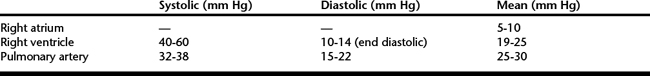
Pulmonary hypertension is characterized by high pulmonary artery pressure (PAP) that leads to impaired right ventricular performance and right-sided heart failure. Normal resting PAP in the adult horse is approximately 32 to 38 mm Hg in systole and 15 to 20 mm Hg in diastole, with a mean pressure of 25 to 30 mm Hg (Table 46-1). During exercise mean PAP correlates with speed of exercise and can reach 120 mm Hg in healthy Thoroughbreds running at maximal speed. There is no clear consensus as to what constitutes pulmonary hypertension in the horse, but systolic pressure greater than 40 mm Hg or mean pressure greater than 35 mm Hg in the resting horse would be in line with most published reports. Primary pulmonary hypertension is idiopathic, whereas secondary pulmonary hypertension results from any process that causes an increase in pulmonary vascular resistance, pulmonary venous pressure, or pulmonary blood flow. Primary idiopathic pulmonary hypertension is a diagnosis of exclusion and has not been characterized in the horse. Secondary pulmonary hypertension has been reported but not fully described in horses with left-sided heart failure resulting from mitral valve regurgitation, with recurrent airway obstruction and with congenital cardiac disease.
PATHOPHYSIOLOGY
The pulmonary circulation is a low-pressure, high-capacity system capable of accommodating large increases in blood flow during exercise. The pulmonary circulation is the major determinant of right ventricular afterload and therefore determines right ventricular output. Pulmonary vascular tone is affected by a number of endothelial-derived and local factors. Nitric oxide and prostacyclin are potent vasodilators, and endothelin-1 and thromboxane A2 are potent vasoconstrictors. An imbalance of these mediators in the pulmonary vasculature leads to vasoconstriction and over time stimulates vascular remodeling. Pulmonary vessels also constrict in response to hypoxia, a reaction meant to direct blood flow to better-ventilated areas of the lung. This is mediated by a variety of actions of hypoxia on pulmonary artery endothelium and smooth muscle cells, including downregulation of endothelial nitric oxide synthase and inhibition of the outward potassium currents triggering pulmonary vascular smooth muscle depolarization.
Three predominant mechanisms are involved in the pathogenesis of pulmonary arterial hypertension: pulmonary venous hypertension, pulmonary overcirculation (volume overload), and increased pulmonary vascular resistance. Pulmonary venous hypertension necessitates a passive rise in pulmonary arterial systolic pressure to maintain a driving force across the vasculature. Volume overload causes shear force injury to the vascular endothelium, which leads to vascular remodeling and luminal narrowing. Increased resistance in the pulmonary vasculature can be induced by hypoxic vasoconstriction, obstructive vascular disease, or parenchymal disorders.
Pulmonary hypertension results in a large mechanical load being placed on the right side of the heart. The right ventricle responds by increasing right ventricular systolic pressure as necessary to preserve cardiac output. As right ventricular filling pressure rises, right ventricular dilation, hypertrophy, ischemia, and right-sided heart failure develop. The ability of the right ventricle to adapt to increased vascular resistance is influenced by how quickly pulmonary arterial pressure rises, and even modest pulmonary hypertension can cause right-sided heart failure if it develops rapidly. Left ventricular failure can also occur as rising right ventricular pressure causes the interventricular septum to bulge and impinge on the left ventricle, decreasing end diastolic volume and cardiac output.
PULMONARY HYPERTENSION AND ACQUIRED HEART DISEASE
Pulmonary hypertension, when secondary to acquired cardiac disease, is usually the result of increased resistance to pulmonary venous drainage and subsequent pulmonary venous hypertension. Pulmonary venous hypertension occurs with diseases that increase left atrial or left ventricular end diastolic pressure or directly compress the pulmonary veins. Severe mitral valve regurgitation is the most common cardiac cause of pulmonary hypertension in the horse. Other acquired cardiac diseases affecting the left side of the heart, such as aortic regurgitation, left ventricular dysfunction, cardiomyopathy, constrictive pericarditis, and left atrial masses, can also cause pulmonary arterial hypertension. More direct obstruction of pulmonary venous drainage can occur in association with less common conditions, such as mediastinal masses and heart-base tumors. Aortocardiac fistula is an acquired cardiac defect that can cause pulmonary hypertension via volume overload similar to many congenital left-to-right shunts. Horses with pulmonary hypertension secondary to acquired heart disease are treated by managing the underlying cardiac disease with the use of appropriate heart failure medications, including positive inotropes, diuretics, and afterload reducers.
PULMONARY HYPERTENSION AND CONGENITAL CARDIAC DISEASE
Pulmonary hypertension is part of the natural history of many types of congenital heart defects and a major determinant of the clinical course and prognosis. Congenital defects with large left-to-right shunts can cause extreme increases in blood flow that exceed the reserve capacity of the pulmonary vasculature when sustained over a long period. In such cases the pulmonary vascular bed is subjected to excessive volume overload and shear forces that damage the vascular endothelium, resulting in intimal fibrosis, medial thickening, and other morphologic changes. The end result is narrowing of the pulmonary vasculature and increased pulmonary vascular resistance. In its most severe form, pulmonary vascular resistance may exceed systemic vascular resistance, resulting in reversal of the shunt from right to left (Eisenmenger’s physiology).
Numerous congenital cardiac defects can result in pulmonary hypertension via left-to-right shunting, including ventricular septal defect, atrial septal defect, aorticopulmonary window, patent ductus arteriosus, and various complex cardiac defects. Anomalous pulmonary venous drainage is a congenital defect that can cause increased PAP by causing pulmonary venous hypertension, similar to left-sided heart disease. At this time, the importance of recognizing the presence or possibility of developing pulmonary arterial hypertension in association with a cardiac defect is associated with formulating a prognosis. In general, horses with medium-to-large left-to-right shunt have a guarded to poor prognosis for athleticism and a reduced life expectancy.
PULMONARY HYPERTENSION AND LUNG DISEASE
Diseases that affect the lungs can elicit pulmonary hypertension in different ways, depending on the underlying disease. Several mechanisms may act in concert to cause increased pulmonary vascular resistance, including hypoxia-induced vasoconstriction, loss of vascular surface area from lung destruction, loss of vascular compliance from hyperinflation-induced vascular compression, and vascular remodeling caused by chronic hypoxic vasoconstriction. Right-sided heart failure secondary to pulmonary disease (cor pulmonale) may develop in either the acute or chronic stages of the pulmonary disease.

Stay updated, free articles. Join our Telegram channel

Full access? Get Clinical Tree

