Chapter 5 Primary Causes of Ear Disease
By definition, otitis externa represents a spectrum of inflammatory changes that occur to the external acoustic canal in response to any insult to the ear canal epithelium. But what actually causes ear diseases? Can putting a bacterium or a yeast organism into a normal ear canal result in disease? Although there may be bacteria or yeasts found in the patient with otitis externa, these organisms are not the cause of the ear disease. The real reason for the ear disease is often overlooked. Primary causes of ear disease are those diseases of the skin that also have a direct effect on the skin lining the ear canal. Cutaneous diseases such as atopy, food hypersensitivity, parasites, foreign bodies, hypothyroidism, and seborrheic diseases frequently result in ear disease.
Trauma
Trauma to the ear canal from injury or inappropriate use of instruments in the ear can lead to primary inflammatory changes within the ear. Hair plucking with curved hemostats can result in traumatic inflammation of the ear canal and resultant infection. A more common reason for trauma to the ear canals is the use of cotton-tipped applicators to clean the ear. Cotton-tipped applicators (Q-tips) and applicators with synthetic materials are very irritating to the epithelium when pushed into an ear canal. Their abrasive effect essentially debrides the layer of surface keratinocytes, which is normally very thin. This results in ulceration of the ear canal and exposure of the dermal elements to the resident bacteria and yeasts in the ear, leading to infection. A Q-tip can also push accumulated material ahead of it as it goes deeper into the decreasing diameter of the ear canal. This may result in a hydraulic effect, with significant pressure and subsequent ruptured eardrum. Cotton-tipped applicators can be used to acquire a cytology sample and as an absorbent material. The cotton tip is laid onto liquid in the ear canal; the cotton absorbs the liquid and is then removed. Q-tips should never be used in a “mopping” motion, going in and out of the ear canal.
Atopic Dermatitis
The ear canal is an invagination of epidermis forming a hollow skin tube in the inside of the head that begins at the eardrum. Pathological mechanisms affecting the skin of the animal have the same effect in the epidermal tube lining the ear canal. Since many diseases found in the ear arise as a result of an underlying skin disease, the veterinarian evaluating the patient with otitis externa should also do a careful evaluation of the pet’s skin to determine the underlying etiology of the ear disease, if possible. Often, proper diagnosis and appropriate treatment of the underlying skin disease diminish the severity of ear disease. The veterinarian should evaluate every otitis externa case for the primary underlying skin disease that has led to the otitis externa. Sometimes something as simple as Otodectes infestation acts as the inciting factor for the patient’s ear disease. Sometimes a much more complicated, multiple-allergen atopic skin disease incites the otitis.
It has been estimated that almost 75% of all canine ear disease is related to atopic dermatitis. Atopy seems to be prevalent in many breeds, giving evidence that this disease may have a genetic origin. Most dogs show clinical signs after the first year of life. Atopic patients probably have a high immunoglobulin E (IgE) response from B-lymphocytes when exposed to individual allergens. IgE antibody binds to mast cells resulting in their degranulation and subsequent release of inflammatory mediators on subsequent exposure to that specific antigen. Often there will be a history of foot licking or chewing, face rubbing, and licking the groin area, in addition to scratching the ears. Many dogs with light-colored coats have the telltale red-orange saliva staining typically found with atopy. Cats may manifest their atopic dermatitis with miliary dermatitis, facial pruritus, or barbering of their hair on the belly and lower legs with their teeth in response to the pruritus. Often dental disease will accompany a pruritic skin disease as hairs get trapped between the teeth and in the gingival sulcus, resulting in gingivitis. Atopic dogs also frequently have secondary bacterial and yeast infections on their skin and in their ears.
Allergies can be seasonal or nonseasonal. In a seasonal allergy, the clinical signs are most intense during the period of high pollen counts and disappear with the reduction of the pollen. Atopic dogs and cats are not allergic to only one type of pollen, mold, or insect, so most allergic ears tend to be nonseasonal, with the severity extending for a long period from the spring to the fall, depending on which pollens, molds, or insects the patient is exposed to. In indoor-only dogs or cats with a nonseasonal history, or in a patient that flares up only during the winter, the atopic otitis may be due to indoor allergens such as house dust mites or molds. It is important to ask the owners about the times of the year in which the ear disease flares up. When clients move or relocate to a new geographical area, changes in pet environment may also cause flareups as the patient is exposed to new antigens.
Atopic dermatitis results from an inflammatory overreaction of the skin to antigenic stimulation. The antigens that remain on the surface of the skin maintain the reaction. In dogs, areas like the feet, face, pinnae, and ventrum have increased mast cell density than other parts of the skin. In cats, the mast cells are concentrated behind the ears and over the dorsum. The areas that are constantly contacting environmental allergens are more often involved in the inflammatory process. Unlike dogs, atopic cats tend to have allergies that also affect the respiratory system (bronchitis and asthma) and the eyes (conjunctivitis) as well as the skin.
Frequent bathing or rinsing of atopic patients with water helps to remove antigens physically from the surface of the skin and hair coat. Ear cleaners, flushes, and wet wipes help to remove antigens from the surface of the pinnae and ear canal. Many cats with ceruminous otitis are atopic, so ear cleaners help remove both the antigens and the cerumen from the ears.
Many patients get rapid relief from their ear disease when the atopic dermatitis is treated with corticosteroid therapy, systemically and/or topically. Most combination otic formulations contain corticosteroids to relieve the inflammation and pruritus in the ear. A new therapy for atopic dermatitis using oral cyclosporine-A–modified capsules (Atopica, Novartis) relieves the pruritic clinical signs without the side effects often seen with corticosteroids. Other dogs and cats get long-term benefit from successful immunotherapy to specific antigens identified through allergy testing. It is important to discuss atopic dermatitis as a cause of ear disease with clients so that they understand the value in pursuing a proper diagnosis. After the otic inflammation resulting from atopic dermatitis is controlled, the ear canal epithelium is not as likely to support bacterial or yeast growth, and the patient can remain comfortable.
Food Allergy
Another common primary cause of canine and feline ear disease is food allergy. Termed “cutaneous adverse food reactions,” many components of this syndrome have a direct effect in the ear canal. Allergies in animals tend to become additive—that is, the severity of the clinical signs increases as the patient is exposed to more and more allergens, such as pollens, molds, insects, and foods. When the total antigen exposure is in excess to the tolerable antigen load, clinical signs develop. Some allergic animals can be controlled with a reduction in allergens. It is not uncommon to do a food trial with a hypoallergenic diet for 2 or 3 months with resultant reduction in otic signs. See Chapter 6 for a discussion of cutaneous adverse food reactions.
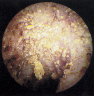
Ear Mites
Ear mites are the most common parasites found in the ear canals of dogs and cats. We are all too familiar with Otodectes mites causing severe damage to the lining of the ear canal, with the resulting “coffee grounds” exudate composed of wax, blood, and epithelial cells. The ear mite has been identified in a number of animal species, both domestic and wild. A nonburrowing psoroptic mite, the ear mite feeds on epithelial cell lymph and blood. They have chewing mouthparts that can cause damage to the epithelium. In dogs and cats, ear mites can cause a severely pruritic parasitic otitis that is commonly associated with a bacterial infection and otitis media.
A unique result of Otodectes infestation in the cat is a systemic hypersensitivity reaction. Known as otodectic mange, this skin disease resembles miliary dermatitis, a papular, crusty eruption found around the neck and head, dorsolumbar area, and inguinal area. When an ear mite–infected cat sleeps with its ear in the flank, the ear mites can leave the ear canal and get on the skin. A similar transfer occurs when an infected cat scratches the ear and the mites get on the paw. Those mites in an ectopic location migrate along the skin and feed. This results in a hypersensitivity reaction to the mite antigens absorbed across the damaged epithelium. Experimentally, infected cats showed an immediate hypersensitivity reaction to an intradermal mite extract. Cats infected for 35 days showed an Arthus (Type III) reaction. Serum precipitating antibodies were noted 45 days after infection. When cats with miliary dermatitis do not respond to systemic steroids, such as methylprednisolone acetate (DepoMedrol, Pfizer), or to flea-control measures, otodectic mange should be considered and the cat should be treated for Otodectes using a systemic acaricide such as ivermectin, fipronil, or sealmectin.
A cat affected with ear mites shakes its head violently and scratches at the ears. Facial abrasions and hair loss may be evident between the lateral canthus of the eyelid and the ear. When examined, the ear canals display the typical reddish-brown to black, dried, crusty exudates. The brown color is presumed to be from dried wax mixed with blood products. On otoscopic examination, the mites can be seen as white insects crawling on the surface of the exudates.
If the otoscope is held very steady, the mite activity increases, because the light arouses the mites and makes them more active. When viewed with a video otoscope (Video Vetscope, MedRx, Inc., Largo, Florida), which has a high magnification and a bright light source, the mites can often be seen in colonies, with thousands of mites scurrying about (Figure 5-1).

Figure 5-1 Otoscopic view of a cat’s ear filled with hundreds of Otodectes mites. The dry, flaky ceruminous exudate is forming a crust in the ear canal.

Stay updated, free articles. Join our Telegram channel

Full access? Get Clinical Tree

