CHAPTER | 16 Pretreatment and Posttreatment Response Images
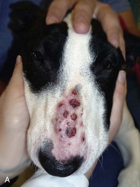
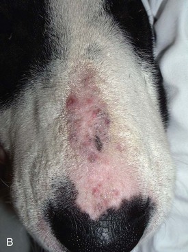
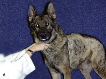
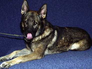
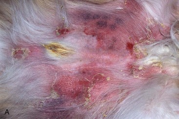
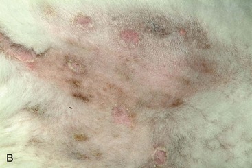
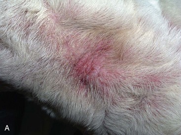
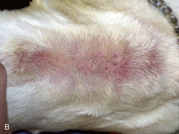
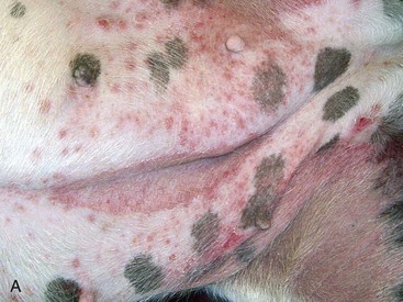
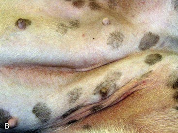
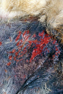
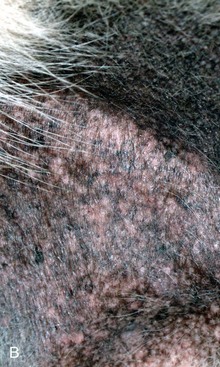
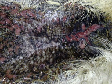
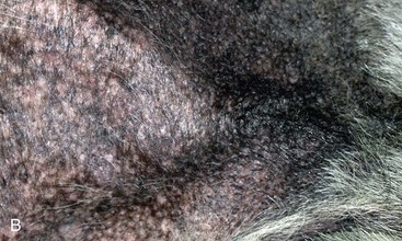
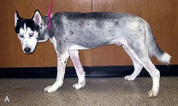
FIGURE 16-7 Pyoderma.
Same dog as in Figure 16-6. A, Erosive lesions caused by the aggressive Staphylococcus infection developed on the abdomen. B, After aggressive antibiotic therapy (high dose, long duration, and optimal frequency), the bacterial folliculitis and furunculosis have resolved.
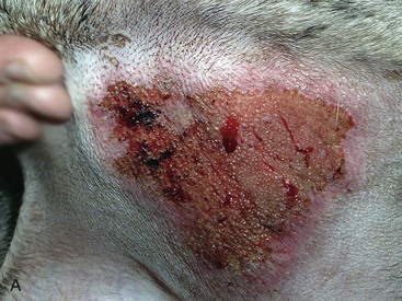
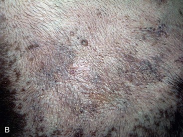
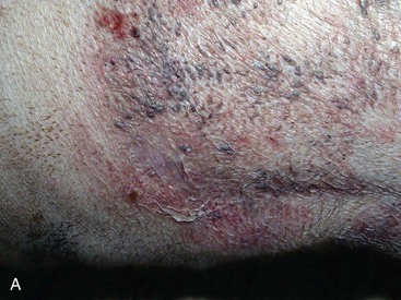
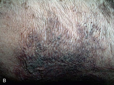
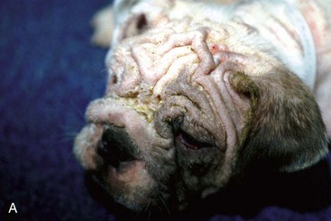
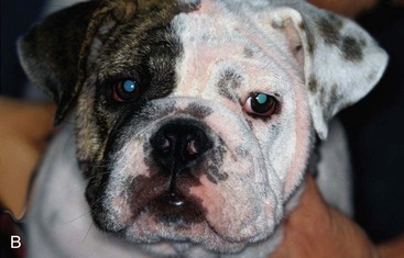
FIGURE 16-9 Nocardiosis.
Same dog as in Figure 16-8. A, This unusual plaque is not typical of the cellulitis lesions associated with nocardiosis. B, After aggressive therapy with trimethoprim-sulfa, the lesions were resolving.
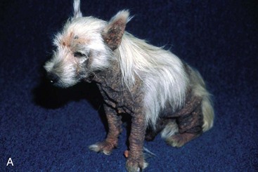
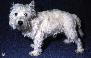
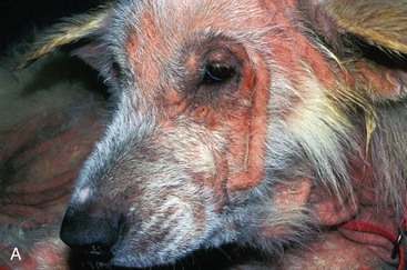
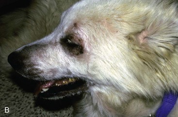
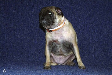
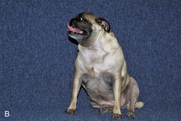
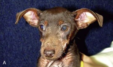
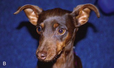
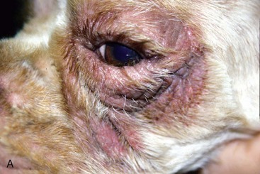
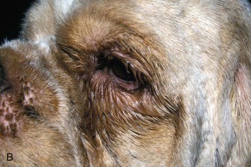
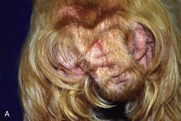
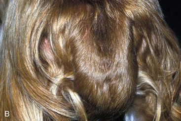
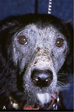
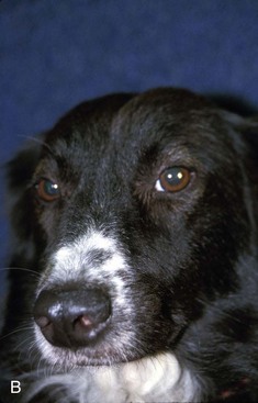
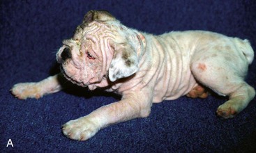
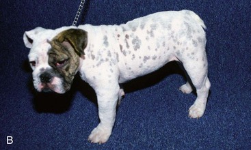
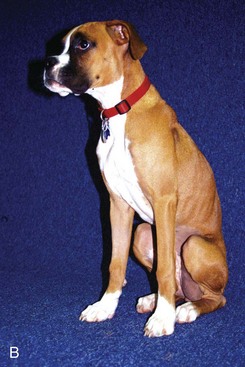
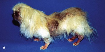
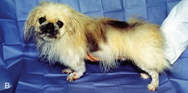
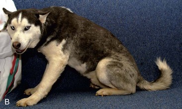
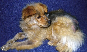
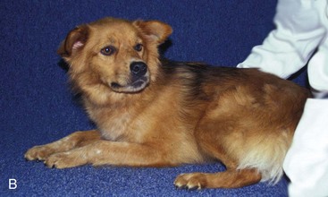
FIGURE 16-15 Malasseziasis.
Same dog as in Figure 16-14. A, Alopecia and lichenification of the tail base caused by secondary yeast dermatitis. Note the similarity to flea allergy dermatitis. B, After several weeks of systemic ketoconazole and topical antifungal therapy, the yeast dermatitis has resolved.
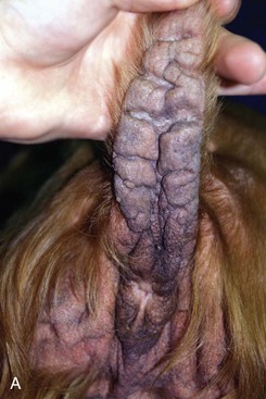
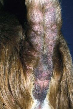
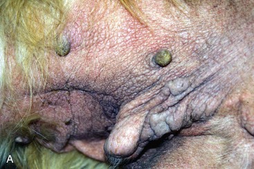
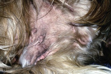
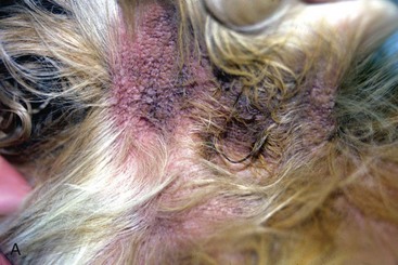
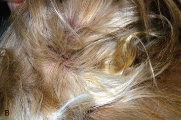
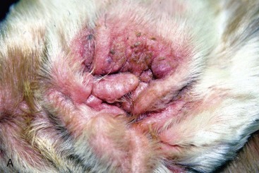
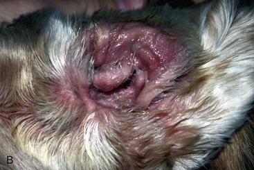
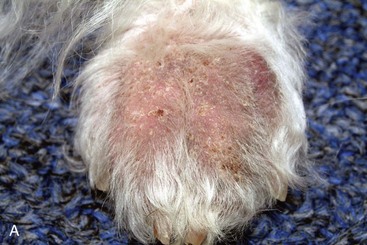
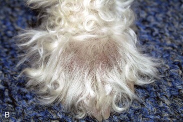
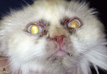
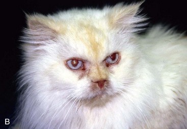
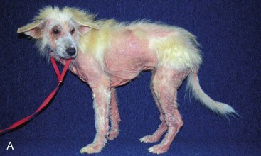
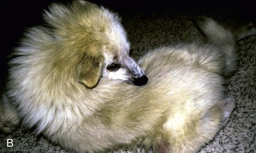
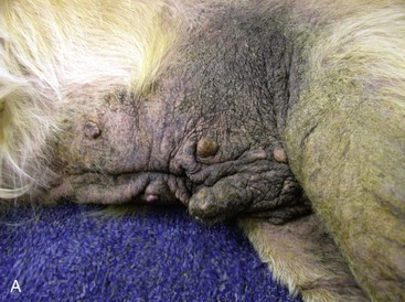
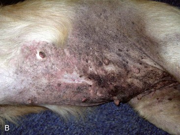
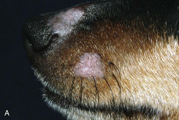
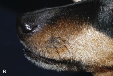
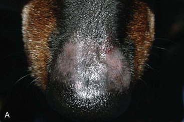
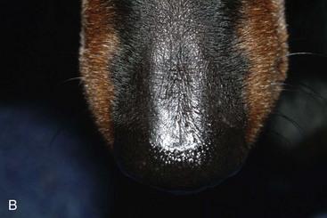
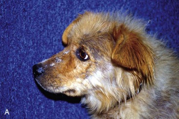
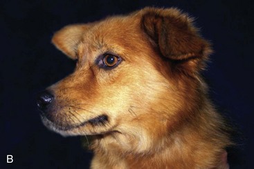
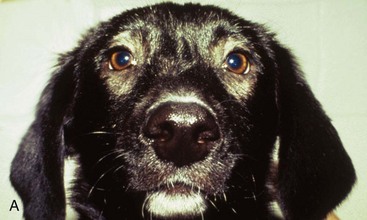
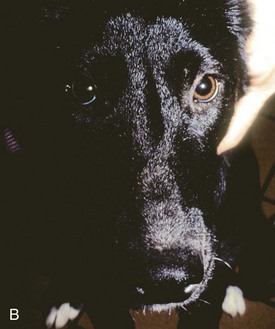
FIGURE 16-25 Dermatophytosis.
Same dog as in Figure 16-24. A, The alopecia and erythema caused by folliculitis affect only the haired portion of the nose, unlike autoimmune skin disease, which would affect nonhaired nasal planum. B, After several weeks of systemic ketoconazole and topical antifungal therapy, the dermatophytosis has resolved.

Stay updated, free articles. Join our Telegram channel

Full access? Get Clinical Tree


