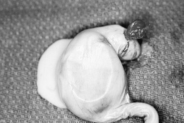
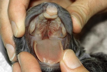
Chapter 219 Conception rates in a normal bitch can exceed 95% with the proper breeding management. Breeding of a fertile queen and tom should result in attainment of pregnancy 70% of the time. The exact number of bitches and queens that undergo embryonic and fetal loss is difficult to determine if pregnancy is suspected but not confirmed before the loss occurs. Methods for pregnancy diagnosis are discussed in Chapter 207. Making a diagnosis of pregnancy loss is also difficult because females may consume the fetuses and show few clinical signs. Pregnancy loss during the second half of gestation often is associated with a hemorrhagic vulvar discharge. Some females that experience fetal death in late gestation do not abort but retain their dead fetuses. Mummification results when fetal death occurs under sterile autolytic conditions. This includes absorption of the placental and fetal fluids and does not occur in the first half of gestation because fetal bone mineralization does not begin until day 40 to 45 of gestation (England, 2006). Mummification can occur in the presence of other live fetuses while maintaining the pregnancy until birth of the live fetuses or all the fetuses can be mummified and be retained past the whelping or queening date. Actual incidence of mummification in the bitch and queen is unknown but it is thought to be low. Retained dead fetuses that are not mummified can become macerated and emphysematous; these require surgical removal. These females may be systemically ill and have a foul vulvar discharge. To determine if a reproduction problem exists, a complete physical examination of the bitch or queen using observation, auscultation, and palpation should be performed. A closer examination of the reproductive system should include vaginal inspection (in bitches) via a speculum or via vaginoscopy to look for discharge or the presence of a fetus, whereas careful vaginal inspection via rectal palpation and digital vulvovaginal inspection and vaginoscopy should be performed in the queen. Abdominal palpation, radiographs, or ultrasonography may be used to confirm pregnancy loss. Confirmation of early embryonic loss in the bitch before implantation is not possible currently in clinical practice, but pregnancy loss after day 21 of gestation may result in a positive relaxin test that persists up to a week after complete pregnancy loss has occurred. Early in gestation, anechoic gestational sacs are visible via ultrasound imaging. In the queen, uterine segmental swellings associated with gestational sacs, fetal poles, heartbeat, and fetal membranes may be seen as early as days 11, 15, 16, and 21, respectively, using ultrasound imaging (Davidson, Nyland, and Tsutsui, 1986). If the gestational sacs fail to develop embryonic structures over time, this confirms pregnancy loss. Later in gestation, fetal death can be confirmed by the absence of a heartbeat and fetal movement during the last 7 to 14 days of pregnancy, which can be confirmed by ultrasound if necessary. Radiographic imaging also can be used to look for signs of fetal death, assess presence and location of remaining fetuses, and estimate the stage of gestation and size of the remaining fetuses. A final form of pregnancy loss is the stillborn. This is well recognized and much easier to confirm. Stillbirths may represent an average of 13% of total kitten births. The mammary glands should be examined for stage of development and presence of colostrum or milk. The primiparous queen tends to develop earlier than the multiparous queen, which can be confusing to the cat owner, making the client concerned about pregnancy loss or undetected abortion. Any fetal, placental, or neonatal materials available should be inspected closely and handled aseptically to allow for further diagnostics (e.g., bacterial culture, virus isolation, karyotyping). Any abnormalities should be noted and investigated. 1. No evidence of pregnancy after breeding 2. Uterine changes suggesting disease 4. Resorbed fetuses or placentation sites in an otherwise healthy bitch or queen First and foremost, optimal breeding management should be discussed at length with the owner. The owner should be advised to confirm the pregnancy after the next breeding so that it can be monitored closely. Older bitches and queens may benefit from additional diagnostic testing (e.g., complete blood count, serum biochemistries, urinalysis, thyroid levels [T4/TSH] in the bitch) along with ultrasonographic imaging of the reproductive tract. A single differential diagnostic plan cannot be followed when working through a case of pregnancy loss in the bitch or queen. Pregnancy loss can be caused by maternal disorders that result in the death of developing embryos or fetuses. This may be caused by disease of a single organ system or be multisystemic. Pregnancy loss also can be caused by fetal disorders that cannot be attributed solely to the bitch or queen (Table 219-1). TABLE 219-1 Primary uterine disease that is inflammatory or infectious may be responsible for pregnancy loss or may mimic pregnancy in some cases (Figure 219-1). Cystic endometrial hyperplasia, pyometra, and metritis are types of inflammatory lesions that can be found in the uterus and may be related to early embryonic death. The lesions may be segmental, likely associated with sites of fetal resorption, or the lesions may be more diffuse, involving one horn or the entire uterus. The bitch or queen may show no clinical signs of disease even when the pathology in the uterus is sufficient enough to prohibit implantation and support of the pregnancy. In other cases abdominal distention may be a notable sign. When the cervix is open, a purulent to serosanguineous vulvar discharge may be present. Cytology of the discharge would reveal an abundance of degenerate neutrophils with intracellular bacteria, suggestive of a septic condition. Figure 219-1 Photograph of segmental pyometra in a queen that could have been mistaken for normal segmental swelling of the uterus because of pregnancy. Ovarian pedicle is located in the upper right of the photograph. Numerous bacteria, including normal vaginal flora (e.g., Staphylococcus spp., Streptococcus spp., Escherichia coli, Mycoplasma spp., and in the queen Coxiella burnetii) and pathogens (e.g., Brucella spp., Campylobacter spp., group G Streptococcus, Leptospira spp., and Salmonella spp.), have been associated with pregnancy loss in the bitch or queen (Pretzer, 2008). More severe clinical signs such as fever, anorexia, dehydration, collapse, and shock can be seen in the bitch and queen when these bacteria cause pregnancy loss. Viral agents are the most commonly reported infectious causes of abortion in queens and include feline panleukopenia (FPLV), FeLV, FIV, and feline herpesvirus-1 (FHV-1). Some of these agents are thought to produce abortion because of the maternal illness and fever in the (FHV-1) infected queen, whereas others, such as FeLV, FIV, and FPLV, cause placental lesions, early embryonic death, and viremia in the fetus (Verstegen, Dhaliwal, and Verstegen-Onclin, 2008). Viral agents involved in pregnancy loss in the bitch include canine herpesvirus (CHV), canine parvovirus (CPV type 1), and canine distemper virus (CDV). Hematologic and biochemistry abnormalities associated with pregnancy loss can include leukopenia or leukocytosis along with a varying degree of azotemia as well as other abnormalities (see Table 219-1). Ultrasonographic examination of the uterus should assist in diagnosis of pregnancy loss. Segmental areas of inflammation may not compromise the pregnancy but can be a reason for concern. Metritis or pyometra, with or without pregnancy, requires medical or surgical treatment (see Chapter 211). Dystocia must be considered as a cause of pregnancy loss in the bitch and queen (see Chapter 208). Other causes of pregnancy loss include uterine displacement, torsion, and uterine rupture, although these conditions do not occur often. Uterine torsion can occur when one or both horns twist along the long axis or around the opposite horn, or the entire uterine body can rotate around itself. Clinical signs can vary depending on the type of torsion that occurs and range from almost no symptoms to those consistent with an acute abdomen. Severe uterine torsions can obstruct blood supply to the uterus, leading to possible rupture of uterine vessels, shock, and fetal or maternal death. Diagnosis is based on clinical signs, diagnostic imaging, and exploratory surgery. Treatment includes hysterotomy to remove the fetuses and correct the torsion or ovariohysterectomy if the uterus is not salvageable. Uterine rupture can follow a uterine torsion or trauma, although this condition is rare. If fetal circulation is not compromised, this condition may go undiagnosed until dystocia results because of the fetuses failing to enter the birth canal. Fetuses expelled into the peritoneal cavity may die immediately and be resorbed (if before fetal bone mineralization has occurred) or become retained fetal mummies. Maternal peritonitis secondary to the uterine rupture is a possible sequel. Treatment includes surgical correction of the uterine rupture, removal of any retained fetuses, and correction of the uterine torsion, if needed. Failure to maintain the pregnancy may result from insufficient production of progesterone (hypoluteoidism/inadequate luteal phase). Progesterone concentrations greater than 1.5 to 2 ng/ml are thought to be required for pregnancy maintenance. This condition could be suspected in a bitch or queen that suffers from repeated pregnancy loss once infectious and other causes of pregnancy loss have been ruled out, although not definitively documented in the queen (Verstegen, Dhaliwal, and Verstegen-Onclin, 2008). Confirmation requires serial progesterone testing. It has been suggested that giving these bitches supplemental progestogen (type and dose remain a source of debate) after day 40 of gestation until 1 week before anticipated whelping date can prevent pregnancy loss. Owners must be warned that progestogen supplementation can cause masculinization of female offspring (pseudohermaphrodism) (Johnson, Root Kustritz, and Olson, 2001) and male offspring that become cryptorchid (England et al, 2006). In addition a decrease in progesterone concentration can be a normal response to terminate an abnormal pregnancy for either maternal (e.g., placentitis, intrauterine infections) or fetal (e.g., primary abnormality, infection) reasons. Pharmaceutic maintenance of an abnormal pregnancy could result in pyometra, dystocia, and even septicemia of the bitch, so caution is important when offering this option to clients. More than 400 genetic diseases have been reported in dogs that have survived gestation, so it is presumed when these defects are more severe, resorption or abortion occurs. Inbreeding lines of dogs and cats also can play a part in neonatal mortality; mixed-breed dogs have a higher reproductive performance than purebred dogs (Johnson, Root Kustritz, and Olson, 2001). Genetic defects are seen with close inbreeding in the cat and may be responsible for 15% of infertility or pregnancy loss or in abortion of domestic animals, including cats. Owners should be advised about the availability of genetic counseling before the start of breeding so that they are less likely to make choices that produce high-risk pregnancies or offspring with genetic abnormalities. Conditions such as cleft palate, thoracic ectromelia, and anasarca are examples of such developmental defects (Figures 219-2 through 219-4). Figure 219-2 Photograph of a full-term puppy with cleft palate, which is a developmental defect. (Photo courtesy Dr. Lawrence Evans.)
Pregnancy Loss in the Bitch and Queen
Clinicopathologic Findings of Pregnancy Loss
Diagnosis of Disorders Associated with Pregnancy Loss
Condition
Species
Inflammatory
Cystic endometrial hyperplasia
Bitch and queen
Pyometra
Bitch and queen
Metritis
Bitch and queen
Infectious
Bacterial
Staphylococcus spp.
Bitch and queen*
Streptococcus spp.
Bitch only
Escherichia coli
Bitch and queen
Mycoplasma spp.
Bitch and queen†
Coxiella burnetii
Queen only
Brucella spp.
Bitch only
Campylobacter spp.
Bitch and queen*
Group G Streptococcus
Bitch and queen‡
Leptospira spp.
Bitch only
Salmonella spp.
Bitch and queen*
Viruses
Canine herpesvirus (CHV)
Bitch only
Canine parvovirus (type 1)
Bitch only
Canine distemper virus (CDV)
Bitch only
Feline panleukopenia (FPLV)
Queen only
Feline leukemia virus (FeLV)
Queen only
Feline herpesvirus-1 (FHV-1)
Queen only
Feline immunodeficiency virus (FIV)
Queen only
Maternal Disorders
Maternal Polysystemic Disorders: The DAMNITT Approach
Developmental/Genetic.
![]()
Stay updated, free articles. Join our Telegram channel

Full access? Get Clinical Tree


Pregnancy Loss in the Bitch and Queen
Only gold members can continue reading. Log In or Register to continue
