Postpartum Disorders in Companion Animals

Chapter 209


Postpartum Disorders in Companion Animals


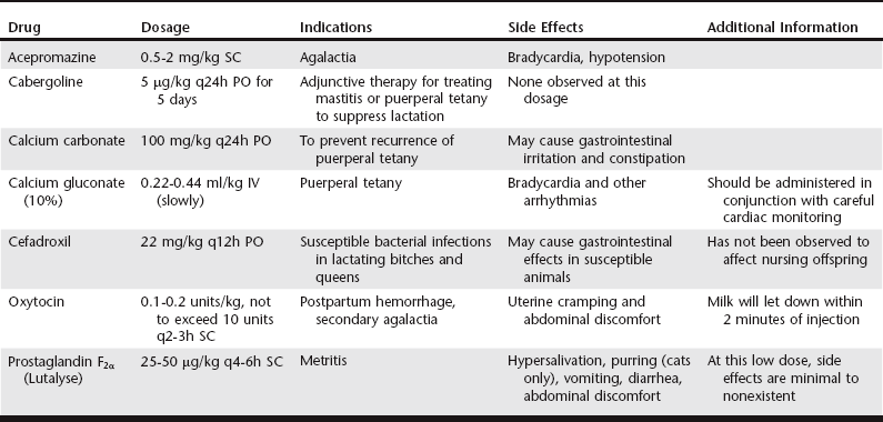
The interval from parturition to weaning, the postpartum period, is typically 6 to 8 weeks in dogs and cats. During the postpartum period, endometrial remodeling at the placental sites occurs, which is histologically complete by 12 weeks after delivery. Postpartum vaginal discharge of lochia, initially dark green in dogs, may persist for 3 to 6 weeks, over which time the discharge changes to reddish brown before it subsides completely. Abnormal events arising during the postpartum period are described in the following paragraphs with a summary of recommended therapies at the end of this chapter (Table 209-1).


Hemorrhage


Scant bleeding after parturition is normal; however, excessive bleeding may indicate uterine or vaginal parturient trauma or may be evidence of an underlying coagulopathy. Vaginoscopy may be useful to identify the source of bleeding (e.g., uterine or vaginal). Intravenous fluid support and blood transfusions may be needed to stabilize the patient, depending on the overall condition of the animal and the total amount of blood lost. When correcting blood volume deficits, clinicians should consider that the normal hematocrit in most bitches and queens at term is 30%, as a result of a hemodilution that occurs during gestation. Although oxytocin (0.1 to 0.2 units/kg, not to exceed 10 units q2-3h SC) may reduce the diameter of the uterine lumen, its efficacy for the treatment of acute postpartum hemorrhage has not been evaluated critically, and ovariohysterectomy often is indicated.


Subinvolution of Placental Sites


Subinvolution of the placental sites (SIPS) involves a delay in the normal process of cytotrophoblast apoptosis (placental degeneration) and endometrial reconstruction. Although the exact etiopathogenesis is unclear, cytotrophoblasts persist within the endometrium instead of degenerating after parturition, preventing endometrial blood vessels from developing thromboses, resulting in persistent endometrial bleeding. Trophoblasts also may invade the myometrium and on rare occasions may penetrate through the serosa, resulting in peritonitis.


SIPS generally occurs in primiparous bitches younger than image years old, with an incidence of 10% to 20% in postpartum bitches. SIPS has never been reported in queens, which is curious given the similarity between the canine and feline placenta. The only clinical sign of SIPS is persistent hemorrhagic (with or without clots) to serosanguineous discharge for more than 6 weeks after delivery. However, SIPS may occur in the absence of any vaginal discharge. A presumptive diagnosis of SIPS is made based upon the clinical history. Bitches with SIPS are afebrile and otherwise healthy, which differentiates SIPS from metritis; however, abnormalities within the bladder or vagina also should be ruled out. Abdominal palpation may identify discrete, firm spheroid enlargements within the uterine horns (approximately 2.5 cm in diameter). Abdominal ultrasonography may identify increased fluid in the uterine horn lumen with prominent placental attachment sites. Identification of multinucleated, basophilic staining trophoblasts with highly vacuolated cytoplasm on cytologic examination of the discharge or histologic examination of a uterine biopsy provides a definitive diagnosis. A cystocentesis with urinanalysis and vaginoscopic examination should rule out the urinary tract and vagina as the source of the discharge.


The clinical management of SIPS must be based on the individual patient and client. Most cases of SIPS are self-limiting, which is fortunate because medical treatment has not proven to be effective. Administration of antibiotics, progestins, or oxytocin does not decrease the duration, amount, or character of the vaginal discharge. Bitches intended for future breeding should be monitored cautiously until the discharge resolves spontaneously. Monitoring includes deliberate daily observation of the bitch’s general appearance by the owner and weekly vaginal cytologic examinations and complete blood cell counts to confirm the absence of metritis or anemia. However, in cases of uterine rupture or severe hemorrhage or if the patient is no longer a valuable breeding animal, ovariohysterectomy is recommended. Normal fertility has been reported in bitches after recovery from SIPS.


Uterine Prolapse


Uterine prolapse is relatively uncommon in the queen and bitch but has been reported in the literature more frequently in queens than bitches. Uterine prolapse can occur during or within a few hours after parturition, involving either one or both uterine horns. Dystocia, delivery of large litters, placental retention, advanced maternal age, and excessive relaxation of the pelvic and perineal region are considered predisposing factors. Examination reveals a firm soft tissue mass protruding from the vagina, which should be differentiated from a vaginal prolapse or neoplasia by palpation of the cervix and clinical appearance. Patients with a uterine prolapse quickly can develop hypotensive or hemorrhagic shock, especially if the ovarian or uterine vessels have been ruptured. While stabilizing the patient, the clinician should cover prolapsed tissues with warm, saline-moistened towels or gauze. After gentle cleaning and copiously lubricating the tissue, manual reduction, with or without an episiotomy, under epidural or general anesthesia should be attempted. Manual reduction may be accomplished with a gloved finger, test tube, or syringe case. Systemic antibiotics are warranted because the risk of metritis after a uterine prolapse is high. Cefadroxil (22 mg/kg q12h PO) is recommended because it has broad-spectrum antibacterial activity and has not been observed to affect nursing puppies or kittens. If the uterine tissue is not viable or the dam is no longer intended for breeding, manual reduction should be followed by an ovariohysterectomy. If the uterus cannot be reduced manually, the urethra should be catheterized, and the uterus may be amputated externally. The amputation procedure involves individual ligation of the uterine arteries, transection, and oversewing of the transected uterine body. A laparotomy should follow amputation to complete the ovariohysterectomy.

< div class='tao-gold-member'>

Stay updated, free articles. Join our Telegram channel

Jul 18, 2016 | Posted by in PHARMACOLOGY, TOXICOLOGY & THERAPEUTICS | Comments Off on Postpartum Disorders in Companion Animals

Full access? Get Clinical Tree

Get Clinical Tree app for offline access