Post-mortem Examination of Rabbits
Post-mortem examinations are an essential part of veterinary practice and, as such, drive forward the evolution of rabbit medicine. In addition to informing and refining diagnoses, they are increasingly requested to provide evidence as part of criminal proceedings. The procedure for post-mortem examination in rabbits is similar to other species. A methodical approach is required (Bivin, 1994). A preprinted checklist of findings is useful, and all stages should be recorded. Initially the carcass is inspected, weighed, palpated and any unusual findings photographed. The carcass should be radiographed before embarking on autopsy. A ventral midline, longitudinal skin incision should be made from the rostral end of the chin to the pubis. The skin can then be incised laterally in the inguinal area and the axilla and reflected laterally. Incision into the midline of the abdominal wall exposes the abdominal organs, which are examined and photographed in situ. Samples of any free fluid should be taken first, and then the organs removed, weighed and portions retained for histopathology, toxicology and bacteriology (paired samples should be taken if the case is likely to be part of legal proceedings). In order to open the thorax, the ribs must be cut; this is easiest below the level of the costochondral junction. The diaphragm can be cut from the ribs and the sternal plate can be reflected rostrally, exposing the heart and lungs. The thoracic viscera should be examined and photographed in situ. Samples of any pleural or pericardial fluid should be taken prior to removal of the heart and lungs. The heart is removed by cutting the great vessels, and can be submitted intact for pathology. The lungs and trachea can be lifted out as one unit, paying careful attention to the larynx and surrounding structures. The head is examined and skinned, paying particular attention to the teeth, ear canals and structures of the cheek and orbit. The brain, nasal cavity and sinuses can be examined by careful removal of the parietal and nasal bone, or the bones of the skull can be preserved for further examination after cleaning. Sample collection for bacteriology and histopathology is important. Many diseases require histopathological confirmation.
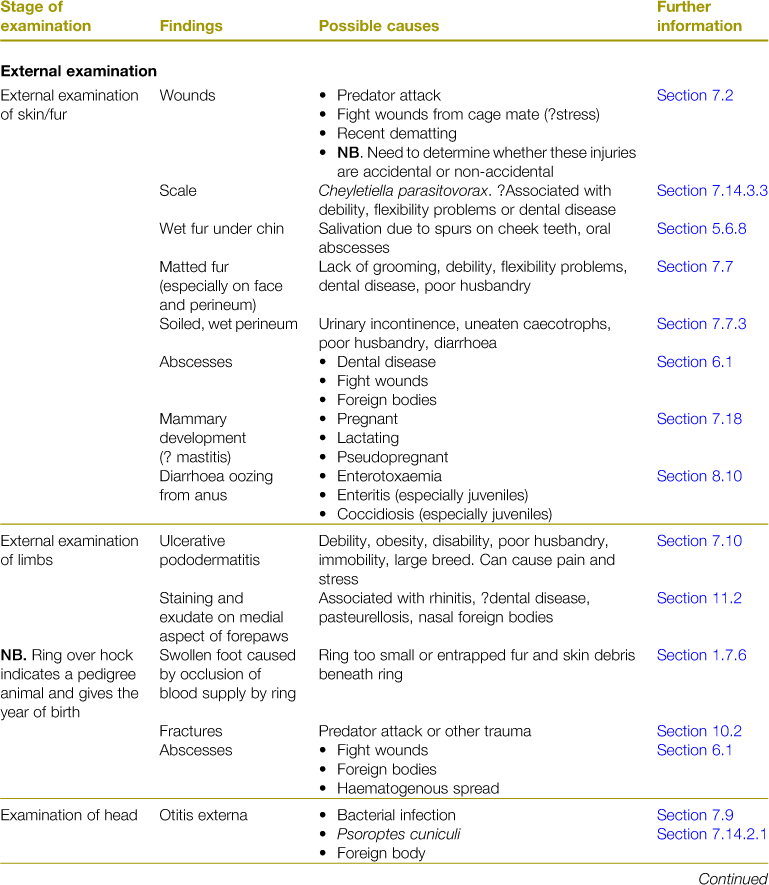
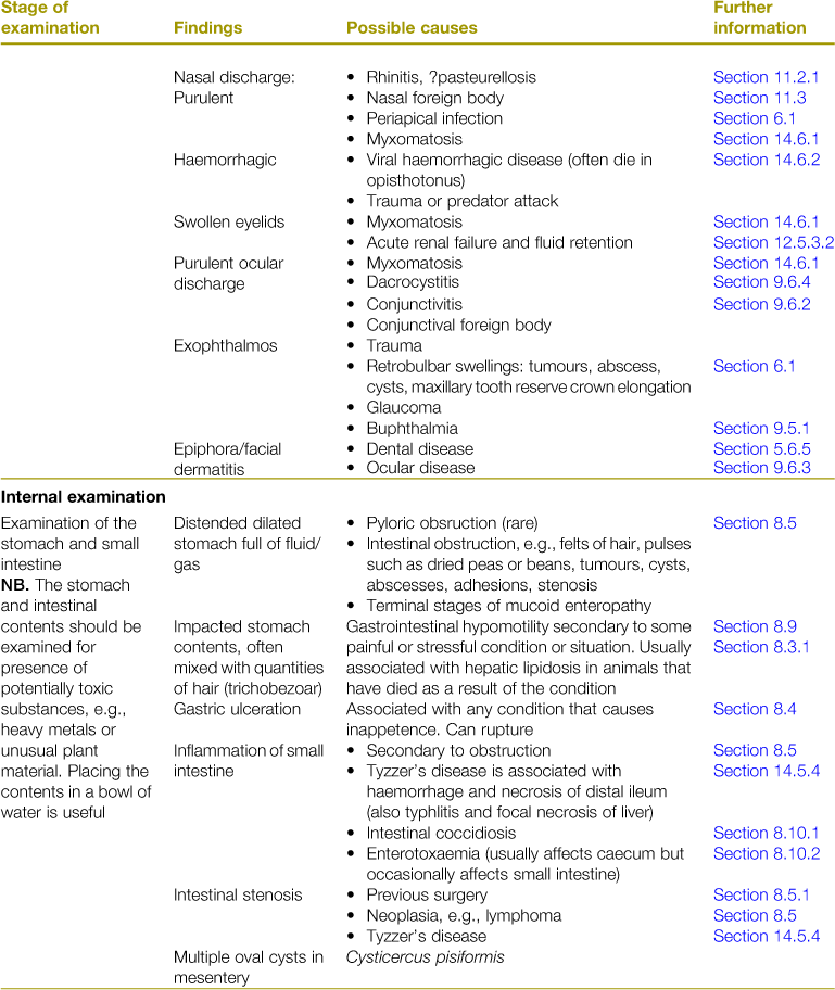
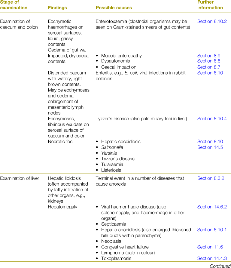
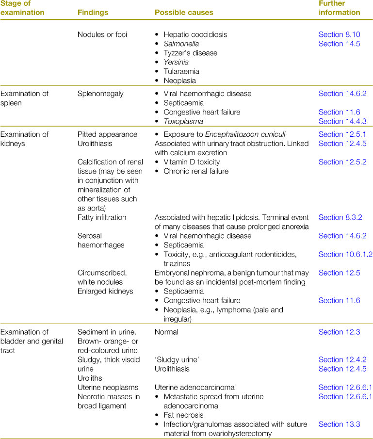
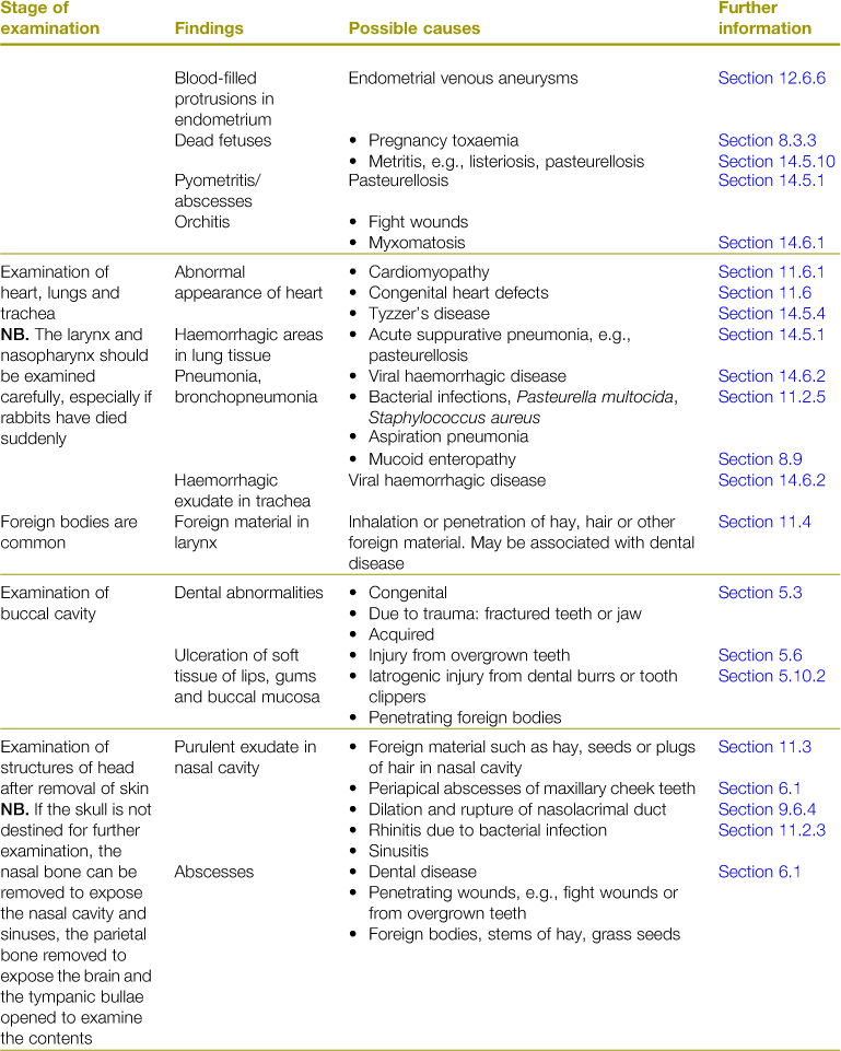
A list of post-mortem findings and possible diagnoses is given in Table 15.1. Some features of post-mortem examination of rabbits are given in Box 15.1. Normal anatomy is illustrated by Barone et al. (1973).
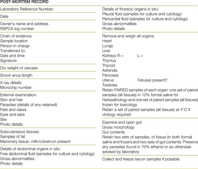
Where a post-mortem is part of the evidence for a legal case, veterinary surgeons should first consider whether they are qualified to undertake the post-mortem examination themselves, and if not, whether they should refer the case to a suitable pathology laboratory. In all cases the ‘chain of evidence’ should be recorded (the location of and person who is in possession of all the samples must be recorded; the time of transfer and to whom must be recorded; and the person who receives the samples should sign a receipt for these) and this information should be included in the permanent case notes (Figure 15.1).
< div class='tao-gold-member'>
