CHAPTER 31 Pneumonia Caused by Rhodococcus equi Infection in Foals
Pneumonia caused by Rhodococcus equi infection in foals is a well-known, worldwide problem. Other less common clinical manifestations of R. equi infection in foals include ulcerative enterocolitis, colonic-mesenteric lymphadenopathy, immune-mediated synovitis and uveitis, osteomyelitis, pyogranulomatous dermatitis, brain abscess, immune-mediated anemia, and septic arthritis. Inhalation of contaminated dust particles is thought to be an important route for pneumonic infection of foals. Ingestion of organism is another important route of exposure and immunization but might not lead to hematogenously derived pneumonia unless the foal has multiple exposures to a large number of bacteria. Recent epidemiologic evidence indicates that foals that develop R. equi pneumonia are most commonly infected during the first few days of life, although clinical signs do not develop until foals are 30 to 60 days of age or older (see Chapter 29, Epidemiology of Rhodococcus equi Pneumonia in Foals).

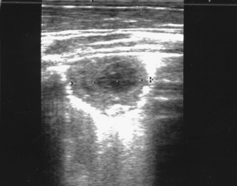
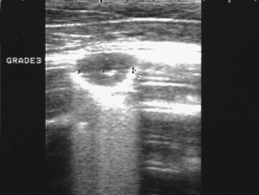
Figure 31-4 Grade 4 pulmonary abscess measuring 3.3 cm in diameter. Notice the hypoechoic center in the lesion.
CLINICAL MANIFESTATIONS
Early clinical signs may include only a slight increase in respiratory rate and mild fever. These signs are often missed, allowing the disease to progress. This results in the signs of respiratory distress appearing to be acute in onset. A small percentage of affected foals may simply be found dead. More commonly, foals are noticed to be in a state of acute respiratory distress and have high fevers (105 to 106° F [40 to 41° C]), even when there were no previous signs or history of respiratory tract disease. Approximately 50% of foals with R. equi
Stay updated, free articles. Join our Telegram channel

Full access? Get Clinical Tree


