Placental Evaluation for Assessment of Foal Problems and Maternal Reproductive Health

CHAPTER 186 Placental Evaluation for Assessment of Foal Problems and Maternal Reproductive Health


Placental incompetence is an important cause of abortion in the mare. Placental adequacy is vital to fetal viability. Normal development and function of the placenta are dependent on maternal health, reproductive competence, and freedom from infection or other diseases. Compromised placental function may result from deficiencies on either the maternal or fetal side of the uteroplacental interface. Systematic and comprehensive placental evaluation gives invaluable insights into the reproductive competency of the mare, the in utero environment of the fetus, and the perinatal health of the foal.


Indications of neonatal compromise attributable to in utero or parturition-related stressors may be subtle; however, prematurity and dysmaturity are often grossly obvious. Other indications of compromise include being underweight for gestational age, abnormal haircoat, or orthopedic abnormalities. Impaired organ and metabolic function result from chronic in utero deprivations but associated changes may not initially be overt. In the initial period following acute hypoxic insult, the foal may appear to have normal behavior and physical examination findings. Aspiration of meconium may not immediately cause detectable pulmonary compromise. Onset of infection and inflammation following vital organ insult is usually progressive but may be preceded by a period in which there are no clinical signs of disease. Timely placental evaluation to detect abnormalities attunes the clinician to the requirement for closer monitoring of the neonate, allowing earlier detection of those subtle variations from normal that indicate neonatal compromise.


The reproductive history of the mare should be considered when the management plan for the impending foaling is formulated. Abnormal vulvar discharge, premature mammary development, or overt loss of colostrum prepartum should alert the clinician to the potential for placental compromise. The resultant fetal distress can also lead to neonatal complications. In situations where premature lactation has occurred, the mammary gland should be assessed for mastitis, and the colostrum should be evaluated for immunoglobulin content at parturition. Prepartum maternal measurements of endocrine function (progestin and total estrogens) are thought to give an insight into the metabolism of the fetoplacental unit, the adequate function of which is paramount to maintaining pregnancy. It has been suggested that increasing progestagen (progesterone metabolite) concentrations seen more than 3 weeks before term, and therefore preceding the normal rise, indicate placentitis or other placental compromise, although no substantive link has been demonstrated with maintenance of pregnancy. Decreased concentrations are considered to indicate fetal compromise. Similarly total estrogen concentrations can be used as an indication of fetal viability, with a normal rise and fall during gestation (between 100 and 320 days) coincident with the size of the fetal gonad.


In a similar vein, ultrasonographic evaluation of the preterm fetus for size, activity level, heart rate, placental dimensions (obtained by both transabdominal and transrectal ultrasound), and fetal fluid characteristics provides an important noninvasive assessment of fetoplacental health. Fetal heart rate varies with fetal activity level, but a resting fetus with a heart rate in excess of 100 beats per minute or less than 50 beats per minute is considered stressed or devitalized, respectively. Increased placental thickness suggests placentitis or vascular compromise. Placental detachment and folding also may be seen during ultrasound examination.


THE NORMAL PLACENTA


The equine placenta is categorized as adeciduate, diffuse, microcotyledonary, and epitheliochorial. These refer to the degree of preservation of maternal tissue, the shape of the attachment area, the method of chorionic attachment to the maternal epithelium, and the degree of invasiveness of the fetal tissues respectively.


No physical attachment exists between the allantoamnion and allantochorion in the horse. The fetus, surrounded by the allantoamnion, floats freely in the allantoic fluid, allowing great mobility in early pregnancy until fetal growth limits movement. This arrangement predisposes the equine fetus to death as a result of umbilical cord torsion.


By convention, vessels leading blood away from the heart are considered arteries, and vessels leading blood toward the heart are considered veins, regardless of their state of oxygenation. By this convention, the umbilical cord consists of two arteries carrying lessoxygenated blood away from the fetal heart, one vein carrying better-oxygenated blood toward the fetal heart (formed from an anastomosis of two proximally), the urachus in the amniotic portion of the cord, and a remnant of the yolk sac. The umbilical cord of the mature fetus normally ruptures soon after parturition a short distance from the body wall. Immaturity of umbilical structures or pathologic changes in this area can lead to excessive blood loss from umbilical or placental vessels or result in abnormal separation allowing a portal of entry for infectious agents.


FACTORS AFFECTING PLACENTAL DEVELOPMENT AND FUNCTION


Placental


Restrictions on placental development and attachment can adversely affect fetal development, with foal weight statistically related to both allantochorionic surface area and allantochorionic weight. The most common cause of diminished placental function is twinning (Figure 186-1). Apposition of the placentas in utero results in a loss of endometrial surface contact, decreasing nutrient and waste exchange ability, leading to chronic deprivation of both fetuses. In twin pregnancies, one fetus is usually confined to one uterine horn only, with the second fetus occupying the remaining horn and uterine body. Deprivation and subsequent death of the more confined fetus can cause placental separation and expulsion of both. Other causes of decreased maternal-fetal exchange include placental edema, chronic placentitis, and premature separation of placenta.
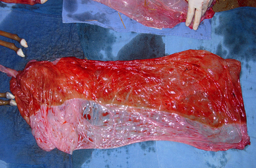


Fetoplacental infections cause up to one third of all abortions. Fetoplacental infections caused by bacteria, equine herpesvirus, fungi, or placentitis of unknown etiology were the most common causes determined in one retrospective study. In another review, pathogens identified most frequently were Streptococcus zooepidemicus, Leptospira spp., Escherichia coli, Pseudomonas aeruginosa, Streptococcus equisimilis, Enterobacter agglomerans, Klebsiella pneumoniae, α-hemolytic Streptococcus, nocardioform actinomycete, and fungi. Mixed bacterial infections were also found. In about one quarter of cases, no causative agent was found. Bacterial placentitis was observed to occur in either of two forms with regard to distribution (focal, diffuse) and time of onset (acute or chronic) (Figure 186-2).
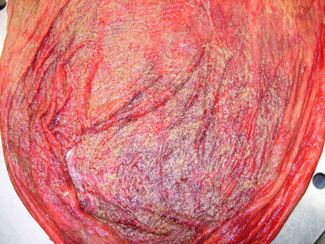


Pathologic changes involving the umbilical cord can restrict blood flow and decrease venous return from the placenta to the fetus. Venous stasis and congestion result in acidemia and anoxia, damaging both the fetus and the placenta. Vital organ function in the fetus may be decreased. Thrombosis of the fetal circulation within the placenta may occur. Decreased perfusion of the chorionic villis results in ischemia and a decrease in functional placental exchange area. A self-reinforcing cycle of fetoplacental pathology can develop.

Stay updated, free articles. Join our Telegram channel

May 28, 2016 | Posted by in EQUINE MEDICINE | Comments Off on Placental Evaluation for Assessment of Foal Problems and Maternal Reproductive Health

Full access? Get Clinical Tree

Get Clinical Tree app for offline access