Pheochromocytoma

Chapter 74 Pheochromocytoma


INTRODUCTION


Pheochromocytoma is a tumor of the chromaffin cells of the adrenal medulla. These cells synthesize, store, and secrete catecholamines in response to sympathetic stimulation (Color Plates 74-1 and 74-2).1 Chromaffin cells are also termed APUD cells, because they are responsible for amine precursor uptake and decarboxylation. Pheochromocytoma may occur alone, or as part of the multiple endocrine neoplasia syndrome. In humans this is a heritable constellation of two or more endocrine neoplasias (or hyperplasia), usually involving the parathyroid and thyroid glands in addition to the adrenal gland.2 Extraadrenal pheochromocytomas (paragangliomas) occur rarely. Most (48% to 80%) of pheochromocytomas in small animals and 30% to 76% in humans are diagnosed on postmortem examination, or as incidental findings on abdominal ultrasonography, and the patient may be clinically asymptomatic.1,3-5 It is thought that pheochromocytomas represent between 0.01% and 0.13% of all canine tumors; however, this number may be low, because the tumor may be benign or nonfunctional and thus not suspected.6 These tumors may be both locally invasive and metastatic.3,5 Most pheochromocytomas in humans secrete norepinephrine (NE) (versus epinephrine), but this has not been studied in dogs or cats.2 It is thought that negative feedback of NE on tyrosine hydroxylase (which converts tyrosine to dopa, leading to synthesis of more NE) does not work normally in the tumor cells, or that the tumor metabolizes NE so quickly that the levels required for negative feedback are never reached.2


CLINICAL SIGNS


In dogs with a pheochromocytoma, approximately 30% to 50% have clinical signs attributable to the tumor. Dogs tend to be older (10 to 12 years),1,5,8 and there is no gender predilection.7,8 Clinical signs may include hypertension, manifestations of hypertension (e.g., blindness from retinal detachment), weakness, collapse, lethargy, vomiting, diarrhea, tachypnea, abdominal distention, syncope, tachyarrhythmias, and/or abdominal pain.5,8 These signs may be sustained or paroxysmal. Because the pheochromocytoma is not innervated like a normal adrenal gland, it is unclear what stimuli cause secretion of catecholamines from the tumor. A Budd-Chiari–like syndrome resulting from tumor invasion and extension up the caudal vena cava has been reported in a dog.9 Approximately 15% to 38% of dogs with a pheochromocytoma have neoplastic invasion of the caudal vena cava; however, clinical signs are not reliably associated with the extent or presence of vena caval invasion.6,10


Concurrent pheochromocytoma and hyperadrenocorticism have been reported in six dogs, and some clinical signs (e.g., panting) may overlap.11 Rupture of pheochromocytomas may result in hemoperitoneum or hemoretroperitoneum.12,13 Dogs may exhibit neurologic deficits or paraparesis secondary to metastatic tumor in the spinal canal, or secondary to aortic thromboembolic disease.5,6,8 Cardiac arrhythmias may include third-degree atrioventricular block, supraventricular tachycardia, or ventricular ectopy.


Of the few cats in the literature with an antemortem diagnoses of a pheochromocytoma, clinical signs consisted of lethargy, vomiting, polyuria, polydipsia, or were associated with systemic hypertension (congestive heart failure and retinal detachment).14-16


DIAGNOSIS


As in humans, most pheochromocytomas in small animals are incidental findings, diagnosed by abdominal imaging or postmortem examination. In some dogs, an abdominal mass may be palpated.5,8


Abdominal radiography may show mineralization in the area of the adrenal glands, or may demonstrate retroperitoneal effusion or an abdominal mass effect associated with the tumor (30% to 50% of cases).5,18 Chest radiographs may show cardiomegaly and pulmonary venous congestion or pulmonary edema secondary to chronic hypertension or tachycardia.1,5 These findings may be confirmed via echocardiography.7 Rarely, metastatic disease may be seen on thoracic radiographs.7,8


Sixty-five to eighty-three percent of pheochromocytomas in dogs are detected via abdominal ultrasonography, making it a useful first-line imaging modality.2 The origin and architecture of the mass, as well as blood flow within the mass and invasion into adjacent structures, may be determined. Pheochromocytomas seem to have a higher likelihood for vena caval invasion than do adrenocortical tumors,16 but ultrasonographically they appear similar to adenocarcinomas.17 It is difficult to determine the cellular origin of an adrenal mass based on ultrasonography, and some masses may be too small for detection by this means.1,2 Invasive pheochromocytomas have been reported to invade not only the vena cava, but also the aorta, renal veins, and hepatic veins.10 Ultrasound-guided biopsies may be obtained, if indicated, but caution should be exercised.


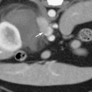
Advanced imaging techniques such as computed tomography (CT) or magnetic resonance imaging (MRI) are very helpful for determining the size of the tumor and the extent of tumor invasion, although these require general anesthesia in the veterinary patient. Nonionic, low-osmolar contrast media is recommended for CT studies to minimize adverse reactions.18 Gadolinium contrast for MRI studies is not contraindicated in patients with a suspected pheochromocytoma.19 CT findings in dogs with pheochromocytoma show a lobulated, irregularly shaped mass associated with the adrenal gland. Areas of decreased intensity are interspersed with highly vascular areas with increased intensity (Figures 74-1 and 74-2).18 MRI may be used to differentiate between histologic types of adrenal tumors.2 Scintigraphy using 123iodinemetaiodobenzylguanidine (an NE analog) or 99mtechnetium-methylene diphosphonate has been used in the dog to identify a pheochromocytoma.20,21 One group used p-[18F]fluorobenzylguanidine to identify tumors in dogs using positron emission tomography.22 These techniques are useful for identifying metastatic tumors.


Stay updated, free articles. Join our Telegram channel

Sep 10, 2016 | Posted by in SMALL ANIMAL | Comments Off on Pheochromocytoma

Full access? Get Clinical Tree

Get Clinical Tree app for offline access