Perpetuating Factors and Treatment of Otitis Externa

Chapter 8 Perpetuating Factors and Treatment of Otitis Externa


Otitis externa is a common malady, occurring in 15% to 20% of dogs and 5% to 7% of cats seen in veterinary practice. Otitis externa is also one of the most frustrating diseases for veterinarians and owners to treat effectively. Treatment regimens vary widely, and a myriad of products containing a variety of ingredients are available for the treatment of ear disease.


Effective treatment of otitis externa often varies from one patient to another; treatments successful in one patient may not help in the next. Each patient and each ear must be considered individually, and treatment regimens must be tailored to the specific case.


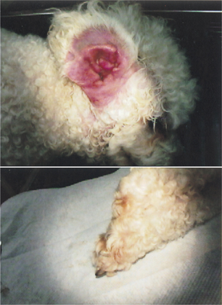
In addition, dermatologic conditions often affect the ear canal, making it susceptible to otitis externa. The ear canal is an invagination of epidermis forming a hollow skin tube inside the head; it begins at the eardrum. Pathologic mechanisms affecting the skin of the animal also affect the epithelial tube lining the ear canal. For example, a dog with atopy may also show inflammation of the ear canal, resulting in redness, swelling, heat, and pain (Figures 8-1 and 8-2). In a cylindrical tube such as the ear canal, inflammation decreases the lumen diameter, tending to reduce both the ventilation and drying of the ear canal. Without ventilation, the humidity level of the ear canal rises, a factor favorable for bacterial growth.


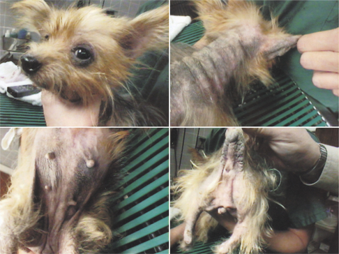
Papules, pustules, crusts, ulcers, and alopecia that may occur on the skin of the trunk may also occur on the skin of the ear canal. Because the ear canal is L-shaped, exudates from these lesions tend to accumulate in the horizontal portion of the ear canal (Figure 8-3). In the treatment of skin disease, shampoo therapy, using a variety of compounds formulated for specific purposes, acts to remove irritating substances and improve healing. In the ear canal, ear flushing solutions containing a variety of ingredients are also used for adjunctive therapy of disease of the ear canal.


New approaches to the medical management of otitis externa in dogs and cats have now been introduced. They include (1) cytologic evaluation of exudates to identify disease processes and determine the type of disease organisms that may be present; (2) flushing products to remove exudates and to disinfect the canal epithelium; and (3) elucidations of pathophysiologic mechanisms showing that otitis externa is a secondary manifestation of underlying skin disease. New combinations of topical medications have been formulated to be effective against bacteria, fungi, and inflammation. The fluoroquinolone antibiotics, injectable ivermectin, and topical fipronil for ear mite infestations have reduced the use of potentially ototoxic antibiotics, oils, and insecticides in the ear canal.


Cytologic Evaluation


The first stop in approaching ear disease is to examine a cytologic preparation of the otic exudates. Examination of a prepared ear smear gives the clinician a starting point for treatment based on the presence of yeasts, cocci, or rod bacteria.


Obtaining samples and preparing a slide to examine constitute a simple procedure that should be a part of the minimum approach to every case of otitis presented to the veterinarian. A sample is obtained with the use of a small cotton-tipped applicator. The swab is inserted through a disinfected otoscope cone positioned near the horizontal canal. The swab is extended beyond the plastic cone, and pressure is applied to the ear canal epithelium as the swab is drawn back through the cone. Every attempt is made to sample from the horizontal canal epithelium only because the vertical canal is often contaminated with a number of commensal organisms unrelated to the ear disease.


The material collected on the swab is rolled onto a clean microscope slide, with the exudates from the left ear on the left part of the slide and the sample from the right ear on the right side. The slide is appropriately labeled with the patient’s name, the date of the collection, and which sample is from which ear. It is then heat fixed and stained with a modified Wright’s blood stain. A drop of slide-mounting medium is placed over the dried, stained material. Then a coverslip is placed on top of the drop, and the glue is allowed to set. The use of slide-mounting medium makes a permanent record of the cytologic characteristics, which can be stored for comparison at subsequent examinations. Alternatively, immersion oil can be smeared along the stained slide and examined.


Examination of the slide under low-power magnification allows an overall view of the cellular debris. High-power examination can help identify and quantify organisms. Cytologic evaluation is very helpful in detecting bacteria and yeasts responsible for secondary infection. Normal commensal bacteria may be found, but in otitis, abnormal increases in numbers of organisms to the point of almost a pure culture, the presence of neutrophils, or both indicate secondary bacterial infection. Often only one ear is affected clinically, but the same organism may be found in the unaffected ear. Sometimes each ear of the same animal has a different organism and needs a different treatment. The severity of the otitis may also be different in the two ears.


When infectious organisms are seen under high-power (400×) magnification, cocci are usually Staphylococcus, and rods are usually Pseudomonas or Proteus. Budding yeasts of Malassezia may be seen individually in the background on a roll smear, but large numbers of yeasts colonizing on exfoliated epithelium indicate secondary yeast infection. Staphylococcus and Malassezia are often found together in the same ear, and there is evidence to suggest that Malassezia growth is stimulated by Staphylococcus.


Bacterial culture and sensitivity testing of exudates may be useful in cases of resistant otitis externa. Many organisms have developed resistance to the routinely used antibiotics, and they should be identified. A limitation of bacterial sensitivity testing is that the organism’s sensitivity or resistance is reported by the laboratory on the basis of the minimum inhibitory concentration (MIC) of the antibiotic in the blood required to kill the bacteria. Topical antibiotics can achieve significantly higher concentrations in the ear canal than systemic antibiotics can in the blood. The high topical antibiotic concentration may actually be effective at killing a bacterium that was reported as resistant. Samples for culture should be taken from the horizontal canal if possible, so that contaminant bacteria are not mistaken for the offending organism. Routine culture of exudates in all cases of otitis is often unrewarding and extremely misleading because four or five bacterial isolates are often reported.


Roll-smear cytologic evaluation becomes useful in determining the etiology of cases of otitis externa without secondary infection. Sheets of epithelial cells may indicate neoplasia as the cause of otitis externa, and the presence of numerous intact nonstaining epithelial cells may indicate a seborreic condition. Parasites such as Otodectes and Demodex, which may be present in the ceruminous exudates, may be found on cytologic preparations, but these parasites are better viewed on a direct mineral oil preparation; this type of preparation is made by rolling the ear swab sample in a drop of mineral oil on a slide. Negative findings on cytology may indicate the need for antiinflammatories only, without the antibiotic or antifungal therapy.


Flushing of the Ear Canal


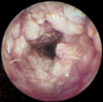
After the class of disease and the type of infection are determined, the next step is to sedate or anesthetize the animal so that a thorough flushing and suctioning of the ear canal can be done. It is imperative that exudates and dried medications that have accumulated in the ear canal be removed so that the canal epithelium itself can be evaluated. Good visualization of the ear canal after flushing helps to ensure that the vertical and horizontal canals are clean and free of debris (Figure 8-4). The efficacy of otic medications is enhanced when they are applied directly onto the cleaned epithelial surface.


Care must be taken in the selection of a flushing agent because so many ear cleaners contain materials that are potentially ototoxic when the eardrum is not intact (see Chapter 17). Prior to using an ear cleaner, the veterinarian should read the label to see whether it can be used when the eardrum is damaged. Many manufacturers are now placing a warning about this issue on their labels.


Mechanical Ear Cleaning


When the ear canal is full of liquid exudates such as pus, warmed saline or warmed, very dilute povidone-iodine solutions (not iodine scrub, which contains detergents) are safe flushing materials to use. Many types of high-pressure irrigation systems are available that can be used to loosen the exudates, but the pressures in such instruments can be very high, necessitating extra caution to avoid rupturing the eardrum. Many of these new irrigation instruments have controls to adjust the flush pressure. Ear curettes, designed to remove debris firmly attached to the epithelium, are useful for scraping the ear canal to dislodge large pieces of wax and epithelial shreds. Curettes are also useful for harvesting cells for cytologic evaluation when a tumor mass is suspected.


After the external ear canal is flushed, it is cleaned with a catheter attached to suction. With good visualization, small pieces of wax, epithelial flakes, and foreign material can be removed. Flushing and suctioning may need to be repeated several times to ensure that the ear canal is thoroughly cleaned.


Cotton-tipped applicators should never be used to clean ears! When a cotton-tipped applicator is pushed into an ear canal full of exudates, the exudate is pushed farther toward the eardrum, making cleaning more difficult and possibly rupturing a weakened eardrum. Cotton swabs are also very irritating to a friable epithelium, and their use may result in painful abrasion or ulceration of the canal epithelium.

Stay updated, free articles. Join our Telegram channel

Sep 10, 2016 | Posted by in SMALL ANIMAL | Comments Off on Perpetuating Factors and Treatment of Otitis Externa

Full access? Get Clinical Tree

Get Clinical Tree app for offline access