CHAPTER 73 Periodontal Disease
Good dental care in the horse includes recognition and treatment of periodontal disease. Although systematic examination and characterization of tissues enables the practitioner to identify the severity of disease, appropriate choice of treatment requires an understanding of the pathogenesis of the disease process. Identification of the stage of the disease process is necessary for the practitioner to provide effective treatment, form a prognosis, and monitor response to treatment.
ANATOMY
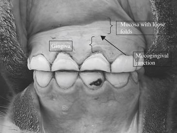
The periodontium is composed of gingiva, periodontal ligament, cementum, and alveolar bone. Gingiva is the part of the oral cavity soft tissue overlying the crowns of unerupted teeth and encircling the necks of erupted teeth. Mucosa is the thin, fragile part of the oral cavity soft tissue that is continuous with the mucous membrane of the cheek, lips, and floor of the mouth. The two tissues meet at the mucogingival junction (Figure 73-1). Periodontal disease is a general term referring to the altered state of the periodontium and encompasses both the active (gingivitis and periodontitis) and resting states of the disease process.
The anatomy of the gingiva can be further specified by location. Marginal gingiva is the unattached terminal edge of the gingiva that surrounds the tooth like a collar. The gingival sulcus is the space around the tooth, bounded by the tooth on one side and the gingiva on the other.Attached gingiva is continuous with marginal gingiva. It is tightly bound to the underlying periosteum and alveolar bone and extends to the relatively loose alveolar mucosa at the mucogingival junction. The interproximal space is occupied by the interdental gingiva (Figure 73-2).
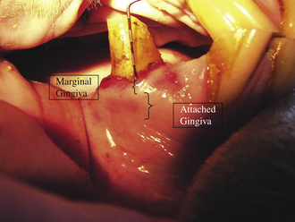
Figure 73-2 Gingival anatomy adjacent to a lower wolf tooth. The periodontal probe is inserted into the gingival sulcus. The tooth borders the gingival sulcus on one side, and the marginal gingiva serves as the outer border. The attached gingiva is secured to the underlying bone. See also Color Plate 7, following p. 352.
Radiographic anatomy of the teeth and supporting structures is similar to that in humans and small animals. Normal structures include the alveolar bone, lamina dura, alveolar crest, and periodontal ligament space. In horses with periodontal disease, changes can be seen in all these structures. Intraoral radiographs provide images of normal and abnormal conditions.
PATHOGENESIS
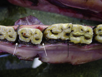
The event that incites the change in bacterial population is decay, which, in humans and small animals, is initiated by the accumulation of plaque and calculus. Although plaque and calculus develop on equine teeth, they rarely lead to periodontitis; the triggering event is stasis of feed material, which decays when it remains lodged in the small depressions around and between teeth (Figure 73-3). The decay process causes inflammation of the gingiva and breaks down cementum and gingival and periodontal attachments. Because equine crowns are covered by cementum, its decay becomes part of the pathologic process. Cemental decay may proceed both apically and interproximally (between the teeth), and destruction of bone and other tissues follows.
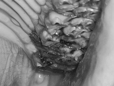
Decreased range of motion also affects the orthodontic movement of teeth (i.e., how teeth move within the arcade). As overlong teeth develop, abnormal mastication forces result. These abnormal forces can cause movement of either of the occlusal pair of teeth and result in an enlarged interproximal space where feed material can accumulate and decay (Figure 73-4). Fortunately, reduction of this malocclusion often results in closure of the interproximal space, particularly in the younger horse.

Figure 73-4 Enlarged interproximal spaces that are a consequence of abnormal orthodontic forces and subsequent shifting of teeth. See also Color Plate 8, following p. 352.
In older horses, these widened spaces can rarely be reduced to normal because the mechanisms that maintain the tight battery of teeth diminish with age. Three processes contribute to enlarged interproximal spaces of aging horses (Figure 73-5): the elastic transseptal fibers of the periodontal ligament, which are compromised as the interdental gingiva suffer damage and fail to keep the teeth in close proximity; overlong teeth block normal mesial drift and prevent maintenance of the close-packed positions of teeth in the masticatory battery; and reduced crown length means teeth have limited ability to erupt. This is especially true of the teeth located distally in the arcade, where the angle of eruption is directed mesially. If the length of these teeth is reduced, they cannot erupt and exert the pushing effect that keeps the rest of the arcade together.