17 Pemphigus foliaceus in a cat
CASE HISTORY
The relevant history in this case was:
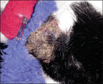
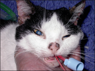
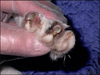
• A 3-month history of crusting lesions initially involving the right pinna with later involvement of the left pinna, face, nasal planum and dorsal trunk. She had also developed a purulent and crusting paronychia. There had been no obvious evidence of pruritus or pain.
• The cat also had a 5-year history of glucocorticoid-responsive, summer seasonal ventral pruritus and symmetrical alopecia over the limbs and tail. Pruritus had persisted despite thorough flea control, but the symptoms had been responsive to corticosteroids. However, corticosteroid therapy had precipitated the onset of diabetes mellitus the previous year and treatment had been withdrawn. The symptoms of diabetes mellitus had resolved on treatment withdrawal.
• She had received routine immunization for feline calici virus, rhinotracheitis and leukaemia 8 months previously. She received regular worming treatment, although not for 2 months prior to the onset of the skin lesions. She received monthly applications of fipronil spot-on.
• At the time of examination, the cat had been treated with enrofloxacin at a dosage of 6 mg/kg s.i.d. with no clinical improvement.
• There had been no response to twice daily applications of a triamcinolone-containing cream to the pinnae.
CLINICAL EXAMINATION
• There was extensive crusting over the pinnae (Fig. 17.1), bridge of the nose (Fig. 17.2), the pre-auricular skin and one focal crusted lesion over the mid dorsum. Removal of the crust revealed shallow, exudative, erosions.
CASE WORK-UP
The following diagnostic tests were performed:
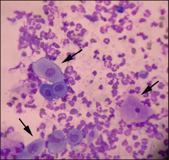
• Cytology of pus from beneath crusted lesions and from the nail folds, which showed numerous neutrophils and acantholytic keratinocytes (Fig. 17.4).
• Histopathological examination of four samples from representative skin lesions, harvested using a 6-mm biopsy punch. There was a mixed superficial perivascular dermatitis, mild to moderate irregular epidermal hyperplasia, and subcorneal pustules/crusts containing mainly non-degenerate polymorphonuclear cells and numerous acantholytic keratinocytes. There was no evidence of bacteria, mites and dermatophytes in the sections examined.
• A blood sample was taken. Routine haematological and biochemical examinations were unremarkable, and FeLV and FIV serology were negative.
AETIOLOGICAL AND PATHOPHYSIOLOGICAL ASPECTS OF PEMPHIGUS FOLIACEUS
Trigger factors
Drug induced: Most cases of pemphigus foliaceus in both cats and dogs appear spontaneously. However, a subset of cases may be drug induced and, in these cases, the disease should resolve when the drug is discontinued. Drug-related pemphigus foliaceus has been reported following administration of cefalexin and trimethoprim-potentiated sulphonamides in dogs, and ampicillin and methimazole in cats.
Stay updated, free articles. Join our Telegram channel

Full access? Get Clinical Tree


