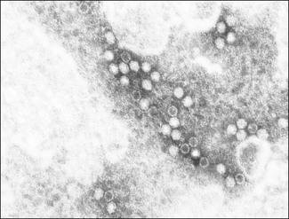
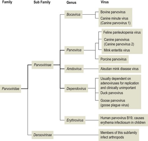
Chapter 45 Parvoviruses (from the Latin word parvus meaning small) are unenveloped, icosahedral in symmetry and possess a linear genome of single-stranded DNA (Fig. 45.1). Their size ranges from 20–26 nm in diameter. The family Parvoviridae is divided into two subfamilies Parvovirinae, which includes viruses of vertebrates, and Densovirinae, members of which infect arthropods (Fig. 45.2). There are five genera within the subfamily Parvovirinae. Viruses of veterinary importance are generally contained within the genus Parvovirus. Two genera have recently been created: Amdovirus and Bocavirus. The genus Erythrovirus contains the human parvovirus B19, which causes erythema infectiosum (‘fifth disease’), a common, self-limiting infection of children. The members of the genus Dependovirus are generally dependent on co-infection with a helper virus, usually an adenovirus, for efficient replication and are found in several animal species. Most dependoviruses are not associated with disease but duck parvovirus and goose parvovirus are not dependent on helper viruses for replication and have been associated with disease in Muscovy ducks and in geese (Derzsy’s disease) respectively. Parvoviruses replicate in the nucleus of host cells. Replication is closely associated with the host cell cycle, requiring the cell to pass through the S-phase and probably involving host DNA polymerases. Parvoviruses are unable to induce resting cells to enter the S-phase and therefore can only replicate in dividing cells. Helper viruses appear to promote cell division rather than becoming directly involved in parvovirus replication. The pathological and clinical manifestations of parvovirus infections reflect their dependence on replicating cells. The single-stranded genome must be converted to a duplex molecule before amplification and RNA transcription of the viral genes can occur. Parvoviruses of veterinary importance are presented in Table 45.1. Table 45.1
Parvoviridae
Virus
Host species
Disease
Feline panleukopenia virus
Domestic and wild cats
Highly contagious systemic disease of cats characterized by fever, depression, vomiting and diarrhoea in weaned kittens. Abortion or cerebellar ataxia in neonates following intrauterine infection
Canine parvovirus
Domestic and wild dogs
Highly contagious systemic disease characterized by depression, vomiting, dysentery
Porcine parvovirus
Pigs
Important cause of stillbirths, mummification, embryonic deaths and infertility (SMEDI syndrome), particularly in gilts
Mink enteritis virus
Mink
Disease of young mink similar to feline panleukopenia
Aleutian mink disease virus
Mink
Immune complex disease, most severe in mink homozygous for pale coat colour. Characterized by persistent viraemia, plasmacytosis and hypergammaglobulinaemia
Goose parvovirus
Geese
Fatal disease of goslings (Derzsy’s disease) characterized by hepatitis, myositis and myocarditis
Canine minute virus
Dogs
Widespread virus, uncertain role in disease
Bovine parvovirus
Cattle
Uncertain role in disease
![]()
Stay updated, free articles. Join our Telegram channel

Full access? Get Clinical Tree


