CHAPTER | 5 Parasitic Skin Disorders


Ixodid Ticks (hard ticks)
Treatment and Prognosis
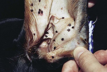
Spinous Ear Tick (Otobius megnini)
Canine Localized Demodicosis
Top Differentials
Differentials include superficial pyoderma, dermatophytosis, and other causes of alopecia.
Treatment and Prognosis
 Ivermectin 0.2–0.6 mg/kg PO every 24 hours is often effective. Initially, ivermectin 0.1 mg/kg PO is administered on day 1, then 0.2 mg/kg PO is administered on day 2, with oral daily increments of 0.1 mg/kg until 0.2–0.6 mg/kg/day is being administered, assuming that no signs of toxicity develop. The cure rate for 0.4 mg/kg/day ivermectin is 85% to 90%.


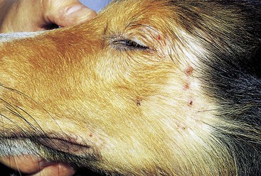
FIGURE 5-3 Canine Localized Demodicosis.
Multiple alopecic papular lesions on the face of an adult Shetland Sheep dog.
(Courtesy D. Angarano.)
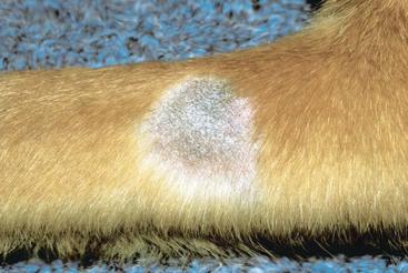
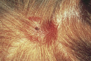
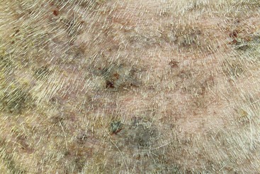
FIGURE 5-4 Canine Localized Demodicosis.
Focal area of alopecia and hyperpigmentation typical of folliculitis.
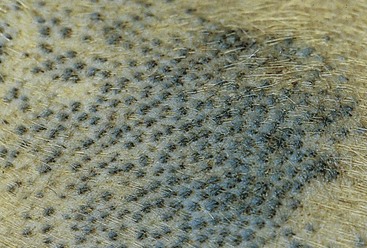
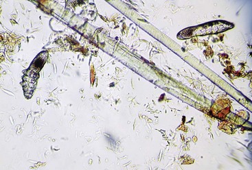
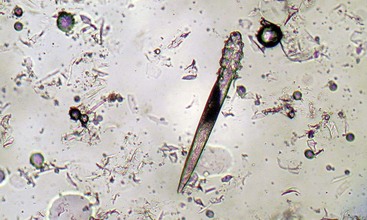
FIGURE 5-6 Canine Localized Demodicosis.
Microscopic image of Demodex mites, as seen with a 10× objective.
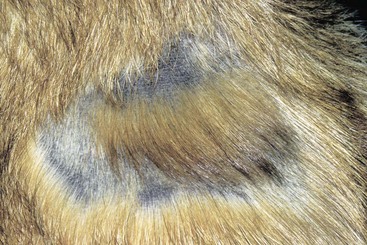
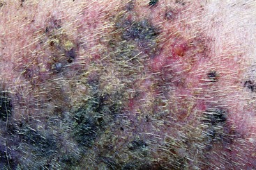
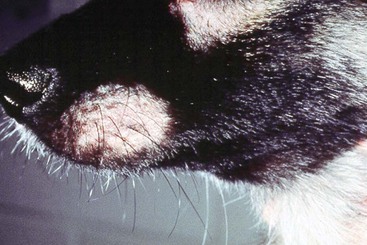
FIGURE 5-9 Canine Localized Demodicosis.
Focal area of alopecia on the muzzle of a young dog.
(Courtesy D. Angarano.)
Canine Generalized Demodicosis
Diagnosis
Treatment and Prognosis
 Ivermectin 0.2–0.6 mg/kg PO every 24 hours is often effective against generalized demodicosis. Initially, ivermectin 0.1 mg/kg PO is administered on day 1, then 0.2 mg/kg PO is administered on day 2, with oral daily increments of 0.1 mg/kg until 0.2–0.6 mg/kg/day is being administered, assuming that no signs of toxicity develop. The cure rate for 0.4 mg/kg/day ivermectin is 85% to 90%.


Steroids are the most common cause of adult-onset demodicosis.
Products containing amitraz tend to be the most toxic usually because of the product vehicle.
Aggressive treatment should be tried for up to 6 months before giving up.
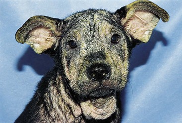
FIGURE 5-11 Canine Generalized Demodicosis.
Generalized alopecia and papules with crusts and scales on the head and neck of a juvenile dog.
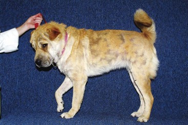
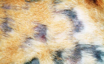
FIGURE 5-13 Canine Generalized Demodicosis.
Close-up of the dog in Figure 5-12. Multifocal areas of alopecia with mild hyperpigmentation are apparent.
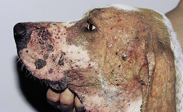
FIGURE 5-14 Canine Generalized Demodicosis.
Diffuse alopecic, erythematous, crusting, papular lesions affecting the entire head and neck.
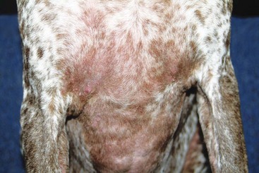
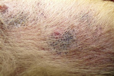
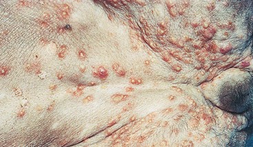
FIGURE 5-17 Close-up of the dog in Figure 5-11. Multiple pustules on the ventral abdomen can be seen.
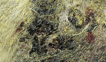
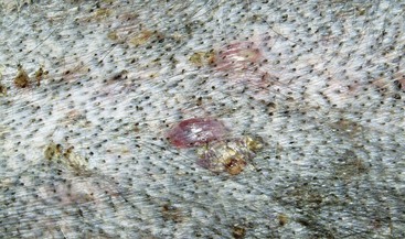
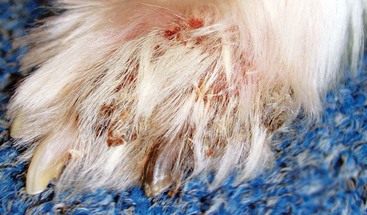
FIGURE 5-20 Canine Generalized Demodicosis.
Matting of the hair associated with an underlying crusting papular dermatitis.
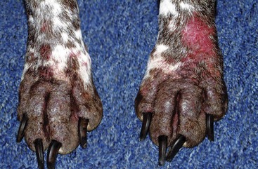
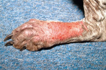
FIGURE 5-22 Canine Generalized Demodicosis.
Alopecia and papular dermatitis with a large erosive lesion.
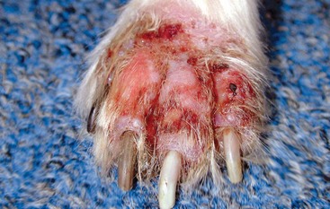
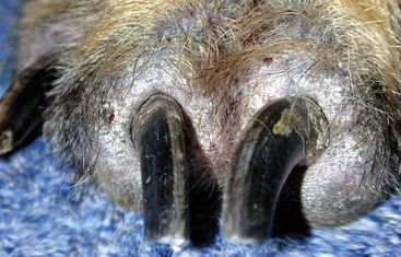
FIGURE 5-24 Canine Generalized Demodicosis.
Alopecia and hyperpigmentation affecting the swollen nail beds (paronychia) of a dog with Demodex.
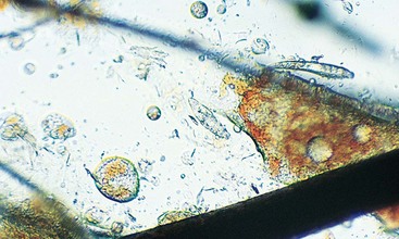
FIGURE 5-25 Canine Generalized Demodicosis.
Microscopic image of Demodex mites, as seen with a 10× objective.
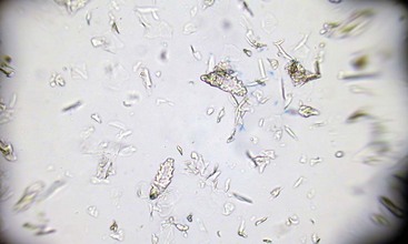
FIGURE 5-26 Canine Generalized Demodicosis.
Microscopic image of Demodex mites, as seen with a 10× objective.
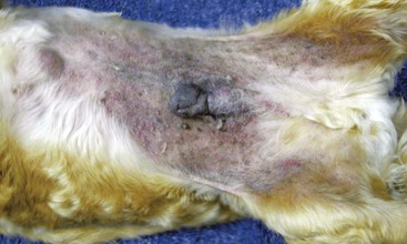
FIGURE 5-27 Canine Generalized Demodicosis.
Diffuse papular dermatitis with hyperpigmentation on the abdomen of an adult Cocker spaniel.
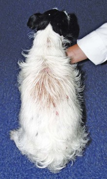
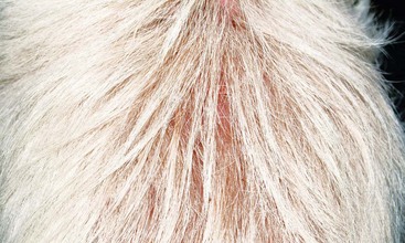
FIGURE 5-29 Canine Generalized Demodicosis.
Same dog as in Figure 5-28. Clumping of the hair is caused by excessive sebaceous secretions.
Feline Demodicosis
Diagnosis
Treatment and Prognosis
 2% to 4% lime sulfur dips applied q 3–7 days for 4 to 8 weeks. Clinical improvement is often observed within 3 to 4 weeks, but therapy should be continued for a total of 6 to 8 weeks to resolve the infection.
 For localized lesions, topical therapies (0.025%–0.03% amitraz solution) may be effective when applied q 24 hours.


