CHAPTER 128 Ossification of the Cartilages of the Foot (Sidebone)
The cartilages of the foot, otherwise known as the ungular or collateral cartilages, are C-shaped structures that are attached to the distal phalanx and extend proximally on the medial and lateral aspect of the foot to just proximal to the coronary band. Sidebone refers to ossification of the cartilages of the foot that usually starts at the cartilage base, at its attachment to the distal phalanx, and progresses proximally. Less commonly, separate centers of ossification may develop that extend proximally or distally and may fuse with the palmar process of the distal phalanx. If ossification originating from a separate center occurs concurrently with ossification from the base of the cartilage, a radiolucent line may exist between the two sites and may persist throughout life. This line may be difficult to differentiate from a fracture. Heterogeneous areas of ossification may also mimic fracture healing. In some horses, there are discrete separate centers of ossification proximally, well separated from ossification further distally. Hypothesized mechanisms of ossification include hereditary disposition, hoof concussion, improper shoeing, hoof imbalance, and poor conformation.
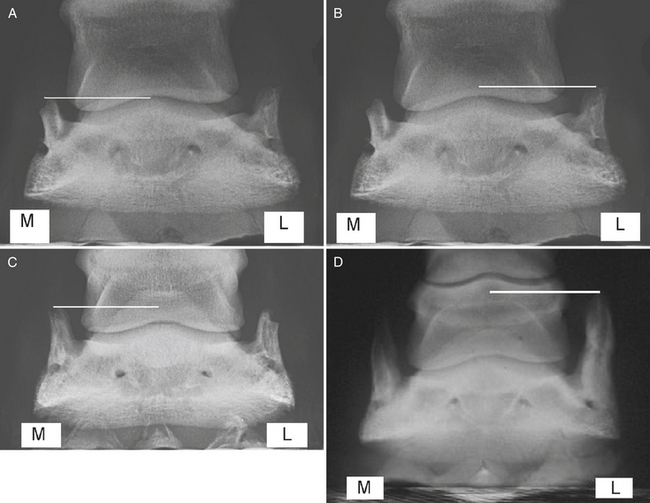
A system for grading ossification of the cartilages of the foot on the basis of a weight-bearing dorsopalmar radiographic view uses the following criteria: grade 0, no ossification; grade 1, minimal ossification at the base of the cartilage; grade 2, mild ossification at the base of the cartilage to the palmar level of the distal interphalangeal joint; grade 3, moderate ossification to the level of the proximal edge of the navicular bone; grade 4, advanced ossification extending clearly above the navicular bone but remaining in the distal half of the middle phalanx; and grade 5, extensive ossification to the level of the proximal half of the middle phalanx (Figure 128-1).
FUNCTIONS OF THE CARTILAGES OF THE FOOT
The distal aspect of the cartilages of the foot contains more venovenous anastomoses than the proximal parts. The greater abundance of venovenous anastomoses at the base of the cartilages may explain why ossification starts distally more often than from a separate center of ossification because this is where most of the energy is first concentrated at the start of dissipation. A recent study has indicated that there is greater radiopharmaceutical uptake (RU) at the base of the cartilages of the foot than further proximally, indicating greater bone modeling. The general symmetry of ossification between left and right feet suggests a hereditary predisposition, which is supported by the estimated high heritability in Finnhorses.


