• How long has the lameness been present? • Has the lameness increased or decreased in severity? • Is the lameness worse in the morning or evening? • Was the limb subjected to some traumatic event? • Does the lameness improve or worsen after a few minutes of activity, such as walking? • Does the lameness change with weather or exercise? • Have other limbs been involved? • Have there been any previous related diagnoses or treatment? With the patient standing, one should observe for weakness, limb trembling, asymmetry of regions of the limbs indicative of muscle atrophy, asymmetry of the head and neck, limb position, and conformation (Figure 10-1). It is common for standing animals to bear less weight on a limb afflicted with lameness. In this situation, the animal may not have the entire foot in contact with the floor. One method to assess the relative amount of weight placed on each limb is for the examiner to place the front feet or the rear feet on the palm of each hand and allow the animal to bear weight on the observer’s hands rather than on the floor. This is a semiquantitative method of assessing weight bearing. Another method to assess weight bearing is to use bathroom scales, or more sophisticated devices described in Chapter 13, under each foot to determine the amount of weight placed on each limb at a stance. In many lamenesses there is also marked muscle atrophy of the affected leg. In some chronic lamenesses there may be abnormal foot pad wear and unequal weight bearing that may be noticeable. Gait evaluation is generally performed after observing the dog rising and at a stance. The patient’s gait is best observed in a large, enclosed exercise area from a distance. In addition, it is beneficial to have an area in which observations may be made with the dog off leash. Gaiting begins with the handler moving the patient first at a walk, then at a trot. The patient should be evaluated while moving directly away from, and then toward the examiner (Figure 10-2). The patient should then be evaluated from the side at a distance (Figure 10-2C). Evaluation at a lope or gallop may be performed if the patient is able, but this gait is less useful for evaluation of most lameness because of the speed of limb motion and the fact that these gaits are asymmetric. It may be helpful to capture the gait with a video camera and then evaluate the gait in slow motion. The degree of lameness may be subjectively evaluated using a lameness scoring system to allow comparison of the lameness over time (Box 10-1). Following observation of the patient for lameness during ambulation, a brief examination is performed while the dog is still standing to assess symmetry and to assess relative muscle mass and the presence of muscle atrophy. Simultaneous palpation of both forelimbs and then both rear limbs will allow the examiner to detect subtle differences between limbs that are normally symmetric (Figure 10-3). Fractures and neoplasia of the musculoskeletal system are usually obvious. Individual regions are evaluated for swelling, abnormal shape, heat, and sensitivity and pain. Evaluation of the rear limbs first may be safer for the examiner while the patient is becoming accustomed to the examiner. The phalanges and metatarsals of the distal limb are evaluated first. The toes are spread apart to evaluate the nail beds, webbing between the toes, and the pads for any infection, trauma, or cracks. The phalanges, interphalangeal joints, metatarsal bones, and metatarsophalangeal joints are individually palpated and assessed for pain, crepitus, swelling, fractures, luxations, or collateral ligament instability (Figure 10-4). The range of motion of the joints is also assessed. The area over the plantar sesamoid bones is palpated for pain and proliferative changes that might be associated with an old fracture or arthritis. Joint stability is further assessed by flexing and extending individual joints and placing varus and valgus stresses on the joints. It is helpful to be familiar with the origin and insertion of the interphalangeal ligaments to properly evaluate instability (Box 10-2). The tarsus is a complex series of joints. It is first assessed for joint effusion and instability. Palpation of the dorsal aspect of the tarsocrural joint is especially rewarding in cases of joint effusion. The joint is placed through a full range of motion to assess the joint for limitations, crepitus, and pain. The examiner’s finger is used to palpate the tarsal bones, with particular attention to the central, third, and fourth tarsal bones. The tarsus has short and long components of the medial and lateral collateral ligaments that are taut in different degrees of flexion and extension. To assess the long components of the collateral ligaments, valgus and varus stresses are applied while the tarsus is placed in full extension (Figure 10-5). Normally, there should be little varus or valgus movement with the hock extended. The short portions of the collateral ligaments are assessed by placing varus and valgus stresses on the tarsus with the tarsocrural joint flexed to 90 degrees. Assessment of these structures while the hock is flexed is more difficult because there is some normal motion in this position. After stabilizing the metatarsus with one hand and the tuber calcis with the other, dorsal and plantar stresses are placed on the hock to determine if subluxation is present or if there has been damage to the supporting plantar structures. With the stifle maintained in an extended position, the hock is flexed to evaluate the calcaneus and common calcaneal tendon. Normally, there should be little ability to flex the tarsus. Excessive flexion may indicate that there has been a fracture of the calcaneus or damage to the common calcaneal tendon. It is also helpful to palpate the insertion of the tendon on the calcaneus to rule out partial avulsion injuries, which may be manifested as a firm swollen area. It is valuable to compare the suspected limb with the contralateral limb to assess subtle findings. Palpation of the tibia is relatively easy because the medial aspect has little muscle covering. The metaphyseal and diaphyseal regions are palpated for periosteal or bone pain, which might indicate hypertrophic osteodystrophy, panosteitis, fractures, neoplasia, or traumatic periostitis (Figure 10-6). It is helpful to palpate the medial and lateral malleoli distally and the head of the fibula proximally. The patella is assessed for medial or lateral luxation. To test for medial patella luxation, the stifle is extended, the distal limb is internally rotated, and pressure is applied to the lateral aspect of the patella to try to displace it medially (Figure 10-7). While maintaining pressure, the limb is slowly flexed and extended to see if luxation occurs with the limb in a position other than full extension. Medial patella luxation is common in toy and miniature breeds and in larger breeds such as Labrador retrievers. In small dogs, the patella may be difficult to locate if patella ectopia exists. In these cases, the tibial tubercle is located and the path of the patellar ligament is traced proximally until the patella is located. To test for lateral patella luxation, the stifle is placed in extension or slight flexion while the distal limb is externally rotated. Pressure is applied to the medial aspect of the patella, trying to force it laterally. Although medial patella luxation is most common in all breeds, if a lateral patella luxation is present, it is most likely a large dog or a chondrodystrophic breed.
Orthopedic and Neurologic Evaluation
History
Initial Observation
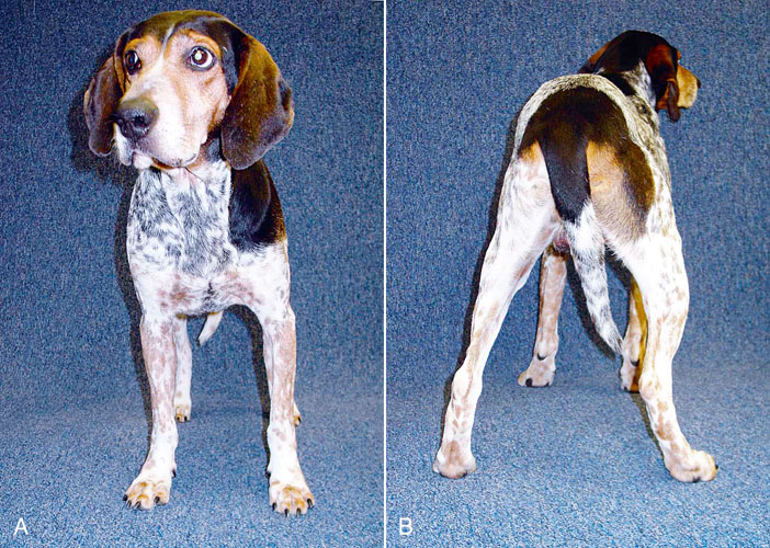
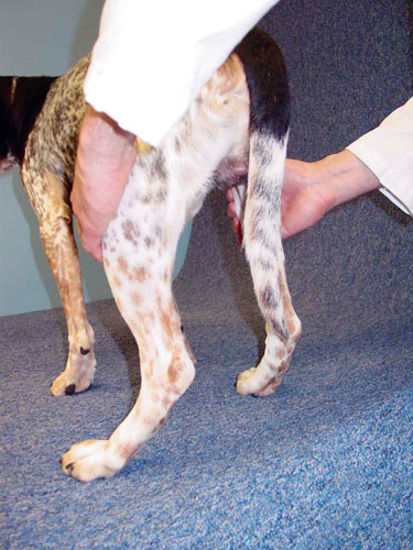
Figure 10-1 The dog should be observed from (A) the front and (B) the rear while standing for evidence of reduced weight bearing on an affected limb, turning the toes out, and not bearing weight symmetrically in comparison with the contralateral limb. Note the reduced weight bearing on the left rear limb indicated by less contact of the metatarsal pad with the ground and the more lateral position of the foot as compared with the other rear limb.
Gait
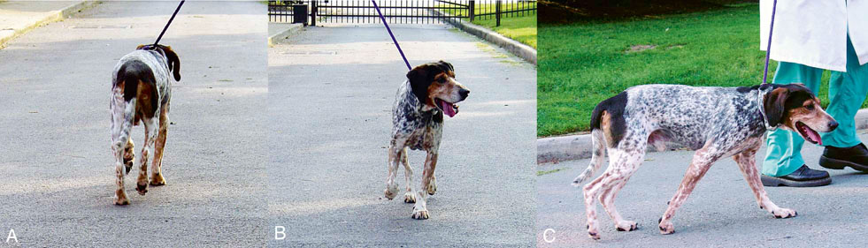
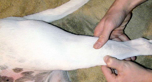
Figure 10-2 The dog should be observed going (A) away from and (B) toward the evaluator at both a walk and a trot. (C) The dog should also be observed from the side.
General Palpation for Symmetry and Atrophy
Orthopedic Examination
Rear Limbs
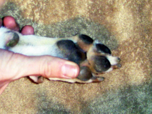
Figure 10-4 The interdigital areas, pads, phalanges, and metatarsal bones should be palpated and the joints placed through a range of motion.
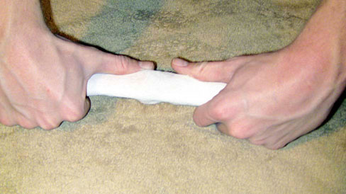
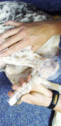
Figure 10-5 The tarsus is evaluated for evidence of medial or lateral collateral ligament damage by applying varus and valgus stresses to the joint.
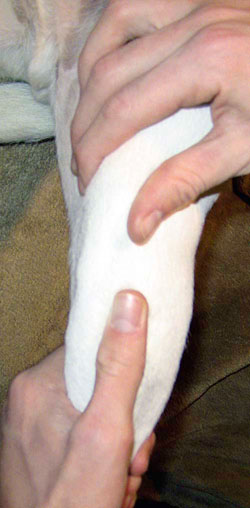
Figure 10-7 To evaluate the stifle for medial patella luxation, the stifle is extended, the tibia is internally rotated, and an attempt is made to push the patella medially out of the trochlear groove.![]()
Stay updated, free articles. Join our Telegram channel

Full access? Get Clinical Tree


Orthopedic and Neurologic Evaluation
Only gold members can continue reading. Log In or Register to continue
