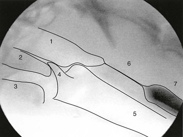
Chapter 121 Oropharyngeal dysphagia (OPD) occurs when the elaborate mechanism of bolus transit from the oral cavity into the esophagus becomes compromised. The swallowing mechanism is complex and involves the action of 31 paired striated muscles and five cranial nerves (sensory and motor fibers of the trigeminal, facial, glossopharyngeal, and vagus nerve, and motor fibers of the hypoglossal nerve), their nuclei in the brainstem, and the swallowing center in the reticular formation of the brainstem. The normal swallowing reflex is a four-stage process, characterized by the oral preparatory phase, oral phase, pharyngeal phase, and esophageal phase. Esophageal and gastroesophageal dysphagias are described elsewhere (see Web Chapter 54). Dysphagia is relatively common in dogs, and the list of possible causes is extensive (Box 121-1). Dysphagia is far less common in cats, and most of the causes of OPD in this species are structural (oral tumors, ulcers, stomatitis). The oral preparatory phase is voluntary and begins as food or liquid enters the mouth. Mastication and lubrication of food are the hallmarks of this phase, as the bolus is modified and prepared for swallowing. Abnormalities of the oral preparatory phase usually are associated with dental disease, xerostomia, weakness of the lips (cranial nerves V and VII), tongue (cranial nerve XII), and cheeks (cranial nerves V and VII). The oral phase of swallowing consists of the muscular events responsible for movement of the bolus from the tongue to the pharynx and is facilitated by the tongue, jaw, and hyoid muscle movements. The pharyngeal phase begins as the bolus reaches the tonsils and is characterized by elevation of the soft palate to prevent the bolus from entering the nasopharynx, elevation and forward movement of the larynx and hyoid, retroflexion of the epiglottis and closure of the vocal folds to close the entrance into the larynx, synchronized contraction of the middle and inferior constrictor muscles of the pharynx, and relaxation of the cricopharyngeus muscle that makes up much of the proximal esophageal sphincter (PES) to allow passage of the bolus into the esophagus (Figure 121-1). Respiration is briefly halted (apneic moment) during the pharyngeal phase. Abnormalities of the pharyngeal phase of swallowing are associated with pharyngeal weakness secondary to neuropathies or myopathies, pharyngeal tumors or foreign bodies, and obstruction of the PES secondary to hypertrophy of the cricopharyngeus muscle. Synchrony between constriction of the pharyngeal muscles and relaxation of the cricopharyngeus muscle is essential to allow passage of the bolus into the esophagus. The esophageal phase is involuntary and begins with the relaxation of the PES and movement of the bolus into the esophagus. Despite the myriad causes of OPD, the pathophysiologic end results fall into one of two interrelated categories: (1) abnormalities of bolus transfer and (2) abnormalities of airway protection. Abnormalities of bolus transfer can be further grouped into those caused by (a) oropharyngeal pump failure (pharyngeal weakness), (b) oropharyngeal and pharyngo-PES discoordination (neuropathies), and (c) pharyngeal outflow obstruction (cricopharyngeal achalasia, tumors of the pharynx, foreign bodies). Figure 121-1 Normal lateral fluoroscopic view of the pharynx at rest. Note that the radiodensity is reversed on fluoroscopic images compared with conventional radiographic images (i.e., air is white, bone is black). 1, Nasopharynx. 2, Soft palate. 3, Base of tongue. 4, Epiglottis. 5, Trachea. 6, Proximal esophageal sphincter. 7, Proximal esophagus with barium in the lumen. (From Pollard RE et al: Quantitative videofluoroscopic evaluation of pharyngeal function in the dog, Vet Radiol Ultrasound 41[5]:409, 2000.) The diagnosis of disorders affecting the oropharyngeal phase of swallowing can be extremely challenging in dogs; however, a history of repetitive swallowing, gagging, and retching associated with meals, nasal regurgitation with meals, swallow-related coughing, falling of food from the mouth during swallowing, and recurrent pneumonia should cause the clinician to suspect OPD (Box 121-2). Dysphagia may be the sole presenting sign in an animal or may be associated with myriad clinical abnormalities. It should be emphasized that OPD may be part of a systemic disease in dogs manifesting signs of dysphagia only, which underscores the importance of a comprehensive systemic evaluation in affected animals. The assessment of dogs with signs of OPD encompasses multiple dimensions that include (1) review of the signalment, (2) review of medication history and inquiry regarding recent anesthesia (Box 121-3), (3) physical examination (prefeeding assessment), (4) neurologic examination, (5) clinical feeding and swallowing evaluation, and (6) laboratory and other testing to provide a basic data set for neurologic evaluation, including imaging studies and endoscopic evaluation of swallowing.
Oropharyngeal Dysphagia
Phases of Swallowing
Diagnostic Approach
< div class='tao-gold-member'>
![]()
Stay updated, free articles. Join our Telegram channel

Full access? Get Clinical Tree


