Orbit and globe – introduction

52 Orbit and globe – introduction


THE ORBIT


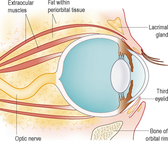
The orbit is the bony fossa which separates the eye from the cranial cavity. Many bones contribute to it – the frontal, lacrimal, maxillary, zygomatic, pterygoid, palatine and sphenoid. The orbit surrounds and protects the globe while providing a route for vessels and nerves to access the eye. In addition to the eye itself, this region contains the extraocular and retrobulbar muscles, optic nerve, lacrimal and nictitans glands, and part of the zygomatic salivary gland, along with the blood vessels and nerves (cranial nerves II, III, IV, V, VI and VII) (Figure 52.1). Connective tissue and fat are also present.


In humans the bony surround is complete, providing excellent protection to the globe from all directions. However, in carnivores there are areas where no bone surrounds the eye – just soft tissue. These areas are mainly ventral to the globe, where the masseter and pterygoid muscles provide the floor of the orbit, and laterally, where the orbital ligament joins the frontal and zygomatic bones. This lack of a complete bony surround is of relevance when considering retrobulbar disease – conditions affecting these muscles of mastication, as well as those in the caudal molars, caudal nasal chambers and zygomatic salivary glands, can all affect the eye due to the close anatomical connection. Thus a dog presenting with a retrobulbar problem needs to have a careful oral examination – a tooth root abscess affecting a molar tooth could easily be contributing to the ocular signs, as could a foreign body which has entered the retrobulbar space via the mouth. Disease processes affecting the frontal and maxillary sinuses can also lead to ocular involvement by local spread of infectious, inflammatory or neoplastic conditions.


The shape of the orbit varies tremendously between canine breeds. Thus in doliocephalic breeds it is a deep concave shape (like an ice cream scoop) while in brachycephalics it can be extremely shallow (more like a teaspoon!). The orbital rim should be palpated as part of the ophthalmic examination and the variation in its position will become apparent. With extreme skull shapes, such as the Pekingese, the orbital rim is markedly posterior to the eyeball itself, and appreciation of this makes it easy to understand how proptosis can occur so easily in such breeds.


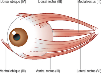
The extraocular muscles are located within the orbit. They are attached to the globe and provide ocular motility. Together with the retrobulbar muscle they form a cone shape with the apex at the optic foramen. They are normal striated muscle. There are six extraocular muscles for each eye: the four rectus muscles (dorsal, ventral, medial and lateral – which pull the eye in their respective directions), together with the dorsal oblique (which pulls the dorsolateral aspect of the globe ventromedially) and the ventral oblique (which pulls the eye mediodorsally) (Figure 52.2). The extraocular muscles are innervated by the oculomotor nerve (cranial nerve III), apart from the lateral rectus muscle which is innervated by the abducens nerve (cranial nerve VI) and the dorsal oblique muscle which is supplied by the trochlear nerve (cranial nerve IV). Thus, if there are abnormalities with the cranial nerves, this can alter both the position of the eye within the socket and affect its movement. These findings can aid with the localization of any lesion and form an important part of neuro-ophthalmology.


Stay updated, free articles. Join our Telegram channel

Sep 10, 2016 | Posted by in SMALL ANIMAL | Comments Off on Orbit and globe – introduction

Full access? Get Clinical Tree

Get Clinical Tree app for offline access