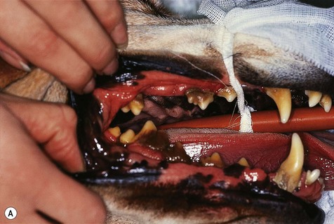
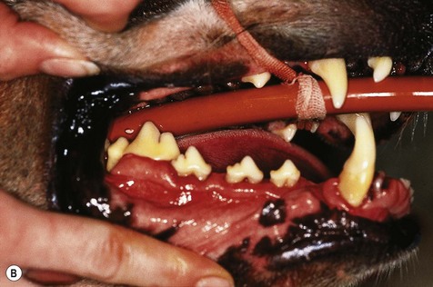
Oral examination and recording

Chapter 6


Oral examination and recording


Oral examination


Examination of the oral cavity is part of every physical examination; however, oral examination in a conscious animal will only give limited information. Definitive oral examination can only be performed under general anesthesia. All detected abnormalities should be recorded. It saves time if one person performs the examination and another individual takes the notes and enters the findings on the dental record.


Conscious examination


Oral examination of a conscious animal is limited to visual inspection and some digital palpation. Gentle technique is essential.


Examination involves assessing not only the oral cavity proper, but also palpation of:


Having looked at the entire face, the mouth is first examined by gently holding the jaws closed and retracting the lips (do not pull on the fur to retract lips) to look at the soft tissues and buccal aspects of the teeth. This is the optimal time to evaluate occlusion. Chapter 5 details the normal occlusal relationships in the dog and cat.


A checklist for evaluation of dental occlusion is shown in Box 6.1.


Finally, the animal is encouraged to open its mouth. One method of achieving this in the dog is to place a thumb and finger on the margin of the alveolar bone caudal to the canine teeth of the upper and lower jaws on one side and with gentle pressure encouraging the animal to open its jaws. Another method, useful for both dogs and cats, is to approach the animal from lateral, one hand is placed over the muzzle and the lips are gently pressed into the oral cavity, while tilting the head slightly upwards. A finger from the other hand is placed on the lower incisors and gentle pressure is exerted. Do not use the fur under the mandible to try to pull the jaw down.


Most animals allow at least a cursory inspection of the oral cavity once the jaws have been opened. The mucous membranes of the oral cavity should be examined as well as the teeth. Apart from color and texture of the mucous membranes, look for evidence of a potential bleeding problem (petechiation, purpura, ecchymoses). In addition, look for vesicle formation and ulceration, which could indicate a vesiculo-bullous disorder, e.g. pemphigus, pemphigoid. Obvious pathology (tooth fracture, gingival recession, advanced furcation exposure) relating to the teeth can be identified. Assess the oropharynx (soft palate, palatoglossal arch, tonsillary crypts, tonsils and fauces) if possible. It is useful to identify any potential problems with endotracheal intubation prior to inducing anesthesia.


Examination under general anesthesia


The oropharynx should be examined prior to endotracheal intubation. Normal anatomic features of the oral cavity need to be identified and inspected. Refreshing your memory on these features from an anatomy textbook is highly recommended. It is only with knowledge of the normal that abnormalities can be identified. A checklist for the oral examination under anesthetic is summarized in Box 6.2.


Periodontium


The periodontium of each tooth needs to be assessed. Examination of the periodontium is not routinely performed in veterinary practice. It is essential to perform a thorough periodontal examination in order to correctly diagnose disease and plan treatment. The procedure for examination of the periodontium is detailed below. Instruments required include:


The following indices and criteria should be evaluated for each tooth:


In animals with large accumulations of dental deposits (plaque and calculus) on the teeth, it may be necessary to remove these to assess periodontal status accurately (Fig. 6.1).


Stay updated, free articles. Join our Telegram channel

Oct 9, 2016 | Posted by in GENERAL | Comments Off on Oral examination and recording

Full access? Get Clinical Tree

Get Clinical Tree app for offline access