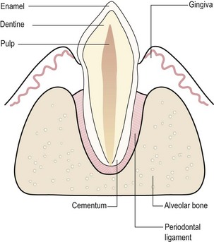
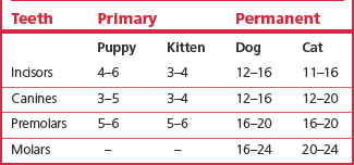
Chapter 4 The dentition of dogs and cats resembles that of man. There are differences in tooth number and shape, but the basic anatomy is similar. The dentition of rodents and lagomorphs is covered in Chapter 14. Each tooth has a crown (above the gum) and one or more roots (below the gum). The bulk of the mature tooth is composed of dentine, which is covered by enamel on the crown and by cementum on the roots. The centre of the tooth contains the pulp or endodontic system. Figure 4.1 depicts the basic structure of a tooth. The crowns of dog and cat teeth have a more tapered shape with sharp cutting edges and fewer chewing surfaces than human teeth. Also, the teeth are spaced further apart and, where there is contact between teeth, the contact area is smaller and not as tight. Humans, dogs and cats are diphyodont, i.e. primary (deciduous) teeth are followed by a permanent dentition. Dental formulae describe the type and number of teeth in each quadrant of the oral cavity. ‘I’ represents incisor teeth, ‘C’ represents canine teeth, ‘P’ represents premolars and ‘M’ represents molars. The respective dental formulae of the primary and permanent dentitions of dog and cat are shown in Box 4.1. Although enamel formation is completed by the time the tooth erupts, dentine production is just beginning. Moreover, root development, i.e. growth in length and formation of a root apex, is by no means complete at the time of eruption. Figure 4.2 depicts maturation of a permanent tooth following eruption. Fig. 4.2 Maturation of a permanent canine tooth after eruption The primary teeth start forming in utero and erupt between 3 and 12 weeks of age. The permanent crowns start forming at or shortly after birth and mineralization of the crowns is complete by around 11 weeks of age. Resorption and exfoliation of the primary teeth and replacement by the permanent dentition occurs between 3 and 7 months of age in the dog and between 3 and 5 months of age in the cat. Once the crowns of the permanent teeth have erupted, root development continues for several months. The approximate ages when teeth erupt in dogs and cats are shown in Table 4.1. Enamel is the hardest and most mineralized tissue in the body. It does not have a nerve or a blood supply. The inorganic content of mature enamel amounts to 96–97% of the weight, the remainder being organic material and water (Fejerskov and Thylstrup 1979). The inorganic material consists of calcium hydroxyapatite crystals arranged in an orderly fashion at right angles to the tooth surface. The organic content is made up of soluble and insoluble proteins and peptides. The enamel of dog and cat teeth is thinner than that of human teeth, generally being 0.2 mm thick in the cat and 0.5 mm in dogs, rarely exceeding 1 mm even at the tips of the teeth (Crossley 1995). This compares with a thickness of up to 2.5 mm in humans (Schroeder 1991).
Anatomy of the teeth and periodontium
Introduction
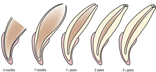
Enamel formation is complete at the time of eruption, while dentine production and root development (root elongation and formation of an apex) are just beginning. The apical foramen of an immature tooth is a single wide opening. As the individual ages, closure of the apex (apexogenesis) occurs by continuous deposition of dentine and cementum until, in mature teeth, the root apex consists of numerous small openings or foramina allowing the passage of blood vessels, lymphatics and nerves.
Anatomy of the teeth
Enamel
![]()
Stay updated, free articles. Join our Telegram channel

Full access? Get Clinical Tree


Veterian Key
Fastest Veterinary Medicine Insight Engine