Chapter 4 Oral-Esophageal Diseases
Initial assessment of the oral cavity can be done with the aid of physical restraint, a mouth gag, and a good light. The gingiva normally is pale pink in color. Although mild gingivitis is very common, association with significant oral disease is unusual. More severe gingivitis, is frequently associated with more serious tooth problems, including tooth loss within 1 or 2 years. With more pronounced redness and edema present diffusely throughout the mouth, deeper tissues may be affected, leading to periodontal disease, with the potential for tooth loss and consequent conditioning problems. If such disease becomes a significant herd problem, regular oral exams are indicated to identify changes in the teeth and to determine if any specific management or dietary changes are likely to help herd performance.1
Palpation of the cheeks can give some insight regarding the health of the cheek teeth. Direct visualization of the cheek teeth requires use of a mouth gag and light source. Even then, a thorough exam is difficult because the animal will continue to chew against the mouth gag. The cheek teeth should be checked for signs of abnormal wear, such as wave mouth, and loss of teeth, which may lead to overgrowth of opposing teeth or food impaction in empty spaces. Molars often will be black because of grass staining, which has no deleterious effect.2 The mandible also should be carefully palpated to detect any bony swelling, which may coincide with tooth root disease.1
Dental care such as floating or clipping abnormally growing teeth may be considered on a case-by-case basis. Implementation of a management program that includes a lot of such dental care probably is best avoided. It can be time-consuming, and each intervention may propagate further imbalance of the dentition, leading to more widespread dental problems within the herd. In treated animals, it also runs the risk of making tooth problems worse if the sensitive pulp cavity is exposed in the shortened tooth. In the case of shortening a cheek tooth overgrown because of a missing opposing tooth, floating is of only short-term benefit, because the missing tooth is the real reason for the problem. The owner must make a decision regarding the management of an animal with sufficiently abnormal teeth that grazing and maintaining body condition have become a problem. This decision centers on supplemental feeding to maintain the animal in production versus culling. Specific considerations include the costs of supplemental feeding as well as replacement costs and availability of confinement facilities to allow such feeding.1
Loss of incisors has important consequences for productivity in most animal management systems that require a lot of grazing. The normal dentition in this area of the mouth should consist of short, closely arrayed incisors. Incisors may become short and peg-like in some young animals pastured on rough grazing. The impaired dentition may become a herd problem over time as a result of decreased grazing efficiency in affected animals. Abnormally long teeth with spaces in between may be a predictor of eventual tooth loss, with consequent significant nutritional problems.
In small ruminant operations, animals with oral-esophageal disease frequently are found to have chronic conditions by the time they are brought to the attention of a veterinarian, largely because they are herd or flock animals: Specific changes in food intake, body condition, and production are not noticed as quickly in the flock as they are in animals that are raised as pets or show specimens. Sheep with poor teeth may have lost a lot of weight before being noticed by owners, because these animals stay with the flock and are observed to eat (although not very efficiently) and move normally.3
1. Spence J., Aitchison G. Clinical aspects of dental disease in sheep. In: Boden E., editor. Sheep and goat practice. London: Baillière Tindall, 1991.
2. Bruere A.N., West D.M. Dental abnormalities. In: The sheep: health, disease and production. Palmerston North, NZ: Foundation for Continuing Education of the New Zealand Veterinary Association; 1993.
3. Scott P.R. Digestive system. In: Sheep medicine. London: Manson Publishing; 2007.
Diagnostic Procedures
Radiography can add important information on conditions of the head, particularly the teeth. Tangential views, typically lateral (Figure 4-1) and dorsoventral projections, often are needed to make an accurate diagnosis and to localize an abnormality. Oblique views are especially helpful in imaging tooth conditions. In oblique views, tooth roots can be evaluated without superimposition of the contralateral arcade. The angle of obliquity is approximately 30 degrees (from lateral), with the x-ray beam directed from ventral to dorsal (Figure 4-2). The affected side should be placed against the cassette. In the 30-degree left ventral–right dorsal view, the right mandible and left maxilla will be profiled on the image. Oftentimes it also is helpful to obtain the opposite oblique view, so that the tooth roots of both arcades can be compared without superimposition of other teeth. Occasionally a 45-degree oblique view can be useful to evaluate the crowns of the teeth without superimposition (Figure 4-3). Tooth root abscesses, broken teeth, skull fractures, and nasal or sinus masses are a few conditions for which skull radiography provides important diagnostic information.
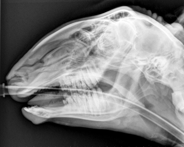
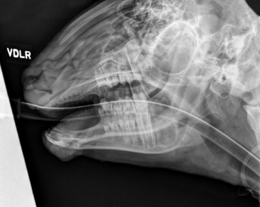
Figure 4-2 Skull radiograph, 30-degree ventromedial–dorsolateral oblique view, of a 6-month-old Suffolk wether, obtained with the animal under general anesthesia. Compare with Figure 4-3.
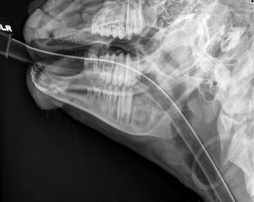
Figure 4-3 Skull radiograph, 45-degree ventromedial–dorsolateral oblique view, of the same animal as in Figure 4-2. Different oblique views may be required for optimal imaging of tooth roots and crowns.
Oral Cavity
The muzzle and oral cavity in sheep and goats are characterized by very mobile lips that are thin relative to those in larger ruminants such as cattle. An obvious philtrum is present in the upper lip. The tongue and palate are smoother than in cattle. The mouth opening is relatively narrow in sheep and goats, compared with that in cattle, making examination of the teeth and oral cavity more difficult. Consistent with findings in all ruminants, the dental pad is located rostral to the palate, where upper incisors are found in other species.1 Small ruminants have three pairs of lower incisors and one pair of lower canine teeth, which look and function just like the incisors. (For the purposes of this discussion, therefore, those canine teeth are referred to as incisors in considering the front teeth as a group.) The dental formula for sheep and goats is 2(Di0/3, Dc0/0, Dp3/3) for deciduous teeth and 2(I0/3, C0/1, P3/3, M3/3) for permanent teeth. Deciduous teeth are in place by the age of 4 weeks in sheep and goats. Aging based on tooth eruption is done by looking at the incisors and canines, which make up the four pairs of rostral mandibular teeth in the small ruminant (Table 4-1). The eruption time for these teeth may vary by 6 months or more, depending mostly on nutrition. The canine is the most unpredictable of these teeth in time of eruption and may even be absent in some mature sheep. One study determined that up to 15.4% of 266 sheep examined lacked either one or both canine teeth, which can interfere with aging by tooth eruption.2
TABLE 4-1 Ages for Permanent Tooth Eruption in Sheep and Goats
| Permanent Tooth | Age at Eruption |
|---|---|
| Incisor 1 | 1 to 1.5 years |
| Incisor 2 | 1.5 to 2 years |
| Incisor 3 | 2.5 to 3 years |
| Incisor 4 | 3.5 to 4 years |
| Premolars | 1.5 to 2 years |
| Molar 1 | 3 months |
| Molar 2 | 9 to 12 months |
| Molar 3 | 1.5 to 2 years |
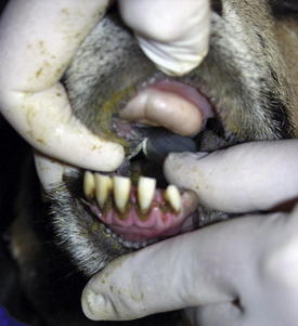
The periodontal ligament holding the incisors is relatively large compared with that in other animals of similar size. This wider ligament allows the movement of the incisors normally seen in ruminants. The normal incisors in sheep and goats are loose enough to be moved a couple of millimeters with gentle digital pressure. The movement minimizes trauma to the cartilaginous dental pad with occlusion and actually aids in cutting plant stuffs when grazing. However, this movability also predisposes small ruminant species to loss of the incisors over time with grazing. Although loss of the incisors can be problematic to the individual affected animal, it may lead to a serious herd management problem with certain rough-grazing pastures if a large percentage of the herd or flock suffers incisor loss, especially at a relatively young age. With loss of incisors, inadequate nutrition related to impaired intake may lead in turn to poor performance by the individual animal or herd3 (Figure 4-4). Loss of incisors is not as dramatic an issue for goats, which are primarily browsers, as opposed to sheep, which graze closely. Goats normally will lose incisors at an older age than is typical for sheep but maintain body condition better than sheep after incisor loss.4
Incisor loss may be due to the use of pasture with sandy soils and consequent wear on teeth from picking up soil during grazing. The teeth are seen to wear on their sides as well as the crown, raising the possibility of other reasons for the excessive wear of the incisors. Acid soils may contribute to this tooth loss, because tooth dentin is demineralized when exposed to (ionized) calcium and phosphate at a pH consistent with some forage and soils.5
Dental health related to ability (or inability) to graze is a very important factor in determining cull rates of sheep. This is true especially in regions or countries in which grazing may be a more important nutritional factor than those in which a lot of supplemental feeding is done. In some management systems it is financially feasible to move older ewes with a bad mouth to supplemental feeding to get another year or two of production, rather than culling and replacing the animals in the flock. The true cost of incisor loss will include increased costs of supplemental feed, lost productive years of ewes, replacement costs for culled ewes, lost production of wool and offspring in ewes with poor dentition, and decreased price of ewes sold with unsound mouths. Broken-mouthed is a term used to describe sheep with one or more missing incisors; gummy describes sheep with all of the incisors missing.6
In contrast with the incisors, the cheek teeth are very stable, with ligamentous support and bone to help grind foodstuffs and cud. Improper wear of cheek teeth may lead to a herd health problem when ewes develop higher-than-normal rates of pregnancy toxemia related to inability to take in enough nutrition to maintain the pregnancy and good body condition. Abnormal wear or loss of cheek teeth may lead to buccal mucosal and gingival abrasions from the remaining teeth as they grow overlong in the absence of opposing teeth, causing trauma to tissues of the oral cavity. The inefficient chewing and pain in the oral cavity will impair nutritional intake, with consequent poor body condition.3
As mentioned earlier, sheep are more adversely affected by lost incisors than goats; however, dental issues that can affect body condition may arise in goats as well: Cheek teeth may wear unevenly, resulting in sharp points that can damage soft tissues of the mouth and make chewing painful. In some animals, tooth root abscesses may form that result in cold sensitivity, leading to decreased water intake. The cheek teeth normally have sharp edges on the lateral aspect of the maxillary teeth and on the medial aspect of the mandibular teeth. If these sharp edges are the likely cause of soft tissue injury, abnormal chewing, and loss of condition, the abnormal points may need to be reduced by filing or cutting. Either dental floats of appropriate size can be used to file the hooks, or the points can be removed by cutting with pliers or Gigli wire. Goats that are having trouble with cheek teeth may be observed to chew on only one side of the mouth, or to drop food while chewing. Some will act hungry but will not eat because of mouth pain. In older goats, oral tumors such as sarcoma, adenosarcoma, osteoma, fibrosarcoma, and fibroma may be the cause of loose teeth, tooth loss, and mouth pain. Cheek tooth root abscesses may cause firm swelling of the area of the affected root. In some cases, at least a temporary response to broad-spectrum antibiotics administered for several weeks may be obtained. Most such abscesses will not heal with use of antibiotics alone, and it often is difficult to financially justify surgical extraction or periapical curettage on any but the most valuable goats.4
Cheek teeth abnormalities are more difficult to determine, because examination and visualization of the cheek teeth can be a challenge. Although gingivitis may lead to abnormal wear and even loss of cheek teeth, the first clinical sign of cheek tooth loss may be loss of body condition. Closer observation may reveal cheek swelling from impacted foodstuffs, or palpation may demonstrate loss of specific teeth. With loss of a tooth, the opposing tooth then grows longer without normal wear. Impact of food on tissue where the tooth was lost or sharp points that form on the remaining tooth may damage soft tissue structures of the cheek, gum, and tongue.7
Sheep with poor dentition that has caused lacerations in the oral cavity will lose body condition, because the oral pain will prevent proper food intake. In some animals, wetting of the jaw with saliva from drooling may be noted. Halitosis also may be noted. Cheek teeth abnormalities frequently cause swellings in the cheek from either oral lesions or impacted foodstuffs. Occasionally the swelling may be retained cud, which can be mistaken for a soft tissue swelling by visual observation alone. Oral examination as already described will make this distinction. Molar teeth abnormalities may manifest as short jerky jaw movements, sometimes with the mouth slightly open. With excessive quidding, fibrous feed may be seen at the commissure of the mouth. Radiographs can be helpful to evaluate cheek teeth; the appropriate oblique view should be obtained to avoid superimposition of tooth roots. The most useful information can be gained if the animal is under general anesthesia for the radiographic study. Palpation of the mandible may detect missing teeth or sharp points on cheek teeth. Cheek tooth abscesses with draining tracts are not frequently seen in sheep.8
The mandible may develop osseous swellings that can be readily discovered on physical examination by palpation of the mandible. Some of these swellings are due to periostitis around tooth roots. Many are of little significance and resolve without treatment. Indeed, some lesions may go unnoticed by the owner. Ones that become too large to ignore or that impair grazing ability probably are due to abscessation of tooth roots. These osseous lesions seldom constitute a herd problem, and although surgical intervention may provide improvement, it often is more costly and time consuming than is reasonable for all but the most valuable of small ruminants. Conflicting results have been achieved with antibiotic therapy, but this usually is worth an attempt to improve the animal’s condition, especially in certain cases, such as that of a pregnant female in which antibiotics may help ensure healthy offspring.3
Fluorosis
The skeletal lesions of fluorosis usually are not apparent until after dental fluorosis is appreciated in the animal. The pathomechanism of the dental abnormalities is fluoride-induced disruption of the normal deposition of mineral in developing teeth. The extent and severity of the fluorotic changes are therefore dependent on the duration of exposure and the age of the animal. The clinical manifestations do not emerge until long after the exposure to fluoride. The clinical signs of abnormal development of dentition include a faster wearing of the teeth that have discolored and chalky and pitted enamel. The dental abnormality observed may be as simple as a groove around a pair of teeth subsequent to short-term exposure of the affected animal to toxic levels of fluoride.7 Details of the pathogenesis and skeletal lesions are presented in Chapter 11.
Stay updated, free articles. Join our Telegram channel

Full access? Get Clinical Tree


