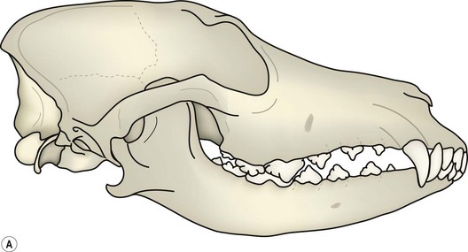
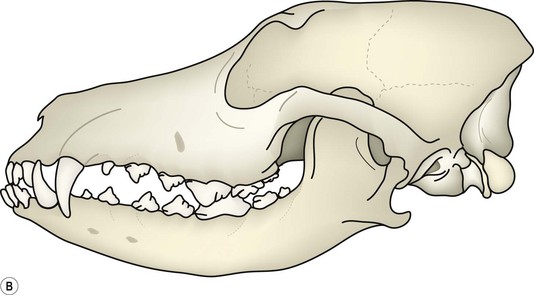
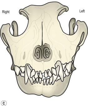
Chapter 5 Malocclusion can result from jaw length and/or width discrepancy (skeletal malocclusion), from tooth malpositioning (dental malocclusion), or a combination of both. The development of the occlusion is determined by both genetic and environmental factors. It is known that jaw length, tooth bud position and tooth size are inherited (Stockard 1941). It is also known that the development of the upper jaw, mandible and teeth are independently regulated genetically (Stockard 1941). Disharmony in the regulation of these structures results in malocclusion. Alteration of jaw growth by hormonal disorder, trauma or functional modification may result in skeletal malocclusion (Hennet and Harvey 1992a). Although tooth bud position is inherited, various events during development and growth may alter the definitive tooth position. It is claimed that at least 50% of malocclusions are acquired and have no genetic cause (Beard 1989; Shipp and Fahrenkrug 1992). There are no data to support such a claim in dogs or cats. Not much research has been done and there are no large epidemiologic studies available. Specific genetic mechanisms regulating malocclusion are unknown. A polygenic mechanism, however, is likely and explains why not all siblings in successive generations are affected by malocclusion to the same degree, if affected at all. With a polygenic mechanism, the severity of clinical signs is linked to the number of defective genes. The most reasonable approach suggested (Hennet and Harvey 1992b; Hennet 1995) to evaluate whether malocclusion is hereditary or acquired is as follows: Fig. 5.1 Scissor bite of the incisor teeth Fig. 5.2 Interdigitation of the canine teeth • The mandibular canine fits into the diastema (space) between the upper 3rd incisor and the upper canine, touching neither. In other words, there should be equal space on either side of the mandibular canine crown. Fig. 5.3 Interdigitation of the premolars Fig. 5.4 Premolar and molar relationships in the dog The incisor and canine occlusion of the adult mesocephalic cat is the same as in the dog. The premolar and molar occlusion differs (Fig. 5.5) from the dog as follows: Fig. 5.5 Premolar and molar relationships in the cat • The most rostral premolar is the maxillary 2nd premolar (the cat lacks the 1st maxillary premolar and the first two mandibular premolars). • The buccal surface of the 1st mandibular molar occludes with the palatal surface of the maxillary 4th premolar. • The maxillary 1st molar is located distopalatal to the maxillary 4th premolar and does not occlude with any other tooth. The cat does not have any teeth with occlusal (chewing) surfaces. Brachycephalic dogs have a shorter than normal upper jaw (Fig. 5.6) and dolichocephalic dogs have a longer than normal upper jaw (Fig. 5.7); in both cases the mandible is not responsible for any rostrocaudal discrepancy. Fig. 5.6 Brachycephalic Fig. 5.7 Dolichocephalic In the mandibular prognathic bite, often called ‘undershot’ (Fig. 5.8), the mandible is longer than the upper jaw and some or all of the mandibular teeth are rostral to their normal position. The degree of malocclusion varies as follows: Fig. 5.8 Mandibular prognathic bite • Normal incisor occlusion, but the mandibular canines touch the upper 3rd incisors and the mandibular premolars are rostrally displaced, which disrupts the ‘pinking shear’ effect. • Level bite: the upper and lower incisors meet at their incisal edges; the lower canines touch the upper 3rd incisors and the mandibular premolars are rostrally displaced. • Reverse scissor bite: the lower incisors are rostral to the upper incisors by 0.5 mm to 5 cm or more; the lower canines may be caudal to but touching the upper 3rd incisors, or may be rostral to the upper 3rd incisors; the mandibular premolars are rostrally displaced to a similar degree. A mandibular brachygnathic bite, often called ‘overshot’, occurs when the mandible is shorter than normal (Fig. 5.9). The degree of malocclusion varies as follows: Fig. 5.9 Mandibular brachygnathic bite • The upper incisors are rostral to the lower incisors by 0.5 mm to 5 cm or more. • The upper canines are caudal to but touching the mandibular canines, level with the lower canines or rostral to the mandibular canines. • The mandibular premolars are caudally displaced relative to the maxillary premolars, disrupting the ‘pinking shear’ effect. The degree of displacement is similar to that of the incisors and canines. A wry bite (Fig. 5.10) occurs if one side of the head grows more than the other side. In its mildest form, a one-sided prognathic or brachygnathic bite develops. In more severe cases, a crooked head and bite develop with a deviated midline. An open bite may also develop in the incisor region so that the affected teeth are displaced vertically and do not occlude. The space between the upper and lower incisors can vary from 0.5 mm to 2 cm.
Occlusion and malocclusion
Introduction
Normal occlusion
Dog
Scissor bite of the incisor teeth (Fig. 5.1)
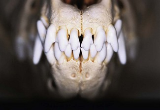
The upper incisors are rostral to the lower with the incisal tips of the mandibular incisors contacting the cingulae of the upper incisors.
Interdigitation of the canine teeth (Fig. 5.2)
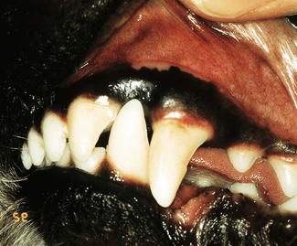
There should be equal space on either side of the mandibular canine crown.
Interdigitation of the premolars (Fig. 5.3)
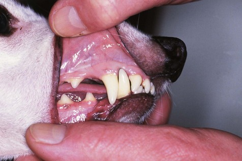
The mandibular 1st premolar should be the most rostral of the premolars.
Premolar and molar relationships (Fig. 5.4)
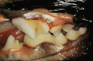
The mesiobuccal surface of the 1st mandibular molar occludes with the palatal surface of the maxillary 4th premolar, and the distal occlusal surface of the mandibular 1st molar occludes with the palatal occlusal surface of the maxillary 1st molar.
Cat
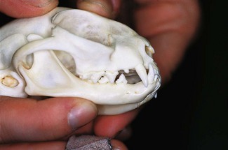
The most rostral premolar is the maxillary 2nd premolar. The buccal surface of the 1st mandibular molar occludes with the palatal surface of the maxillary 4th premolar. The maxillary 1st molar is located distopalatal to the maxillary 4th premolar and does not occlude with any other tooth.
Skeletal malocclusion
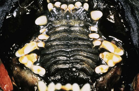
Brachycephalic dogs have a shorter than normal upper jaw. A short jaw results in reduced interdental spaces with rotation and/or overlap of teeth.
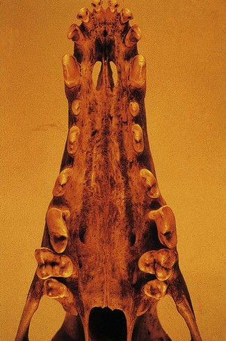
Dolichocephalic dogs have a longer than normal upper jaw. The increased jaw length results in interdental spaces that are wider than normal.
Mandibular prognathic bite
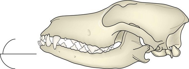
The mandible is longer than the upper jaw. This is normal for brachycephalic breeds.
Mandibular brachygnathic bite
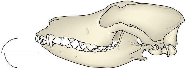
The mandible is too short in relation to the upper jaw. This is normal for dolichocephalic breeds.
Wry bite
![]()
Stay updated, free articles. Join our Telegram channel

Full access? Get Clinical Tree


Occlusion and malocclusion

