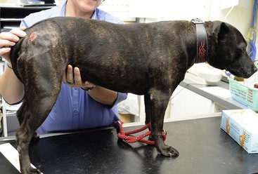
Chapter 104 Canine leproid granuloma (CLG) is a cutaneous or subcutaneous, typically self-limiting nodular mycobacteriosis caused by a novel mycobacterium yet to be fully characterized. The causal organism is distributed worldwide and is common in Australia and Brazil, as well as parts of Europe and the United States (Foley et al, 2002). The route of inoculation is unknown, but biting flies, midges, mosquitoes, or other arthropods may introduce mycobacteria into the host based on the following evidence: the presence of potential vectors around affected dogs, lesions occurring at sites favored by such vectors (e.g., the dorsal fold of the ears), and the prevalence of CLG in short-coated breeds and dogs housed outdoors (Malik et al, 1998). CLG occurs almost exclusively in short-coated breeds, with boxers and boxer crosses remarkably overrepresented. Staffordshire bull terriers and Doberman pinschers also are commonly affected (Malik et al, 2001). The finding of multiple characteristic lesions in typical locations in a short-coated breed suggests the diagnosis of CLG. CLG usually is manifest as single or multiple firm, granulomatous to pyogranulomatous, well-circumscribed nodules in the skin or subcutis (Figure 104-1). Nodules typically are located on the head, particularly the dorsal fold of the ears. Some nodules ulcerate and are usually painless, but may be pruritic when secondary infection with Staphylococcus pseudintermedius occurs. Affected dogs are otherwise healthy. Figure 104-1 A single cutaneous nodule in a Staffordshire terrier with canine leproid granuloma (CLG) and secondary Staphylococcus pseudintermedius infection. The dog made a complete recovery following surgical excision with wide margins. Note the majority of CLG locations are found around the head and ears, but they can occur elsewhere on the body as in this case. (Photo courtesy Anne Fawcett.) PCR testing provides a definitive diagnosis by amplifying regions of the bacterial 16S ribosomal RNA gene using mycobacterium-specific primers. The test is more sensitive when performed on DNA extracted from fresh tissue; however, published PCR protocols generally are successful even when used on paraffin-embedded, formalin-fixed tissue, although false-negative results may occur when contact time with formalin is over 48 hours. Retrospective studies using PCR have demonstrated the presence of microbial DNA (e.g., from mycobacteria and Leishmania spp.) in specimens previously diagnosed as sterile. Recently, one of the authors (JF) has developed a real-time PCR test for the CLG organism that is sensitive and specific and improves the availability and accuracy of molecular diagnostics (Smits et al, 2012).
Nontuberculous Cutaneous Granulomas in Dogs and Cats (Canine Leproid Granuloma and Feline Leprosy Syndrome)
Canine Leproid Granuloma
Causes
Signalment and Clinical Findings

Diagnosis
![]()
Stay updated, free articles. Join our Telegram channel

Full access? Get Clinical Tree


