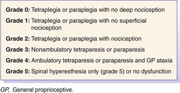
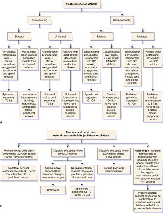
Neurologic Examination and Neuroanatomic Diagnosis

Chapter 26


Neurologic Examination and Neuroanatomic Diagnosis


The past 10 years have been marked by a dramatic increase in the availability of advanced neurodiagnostic imaging for veterinary patients. Magnetic resonance imaging (MRI), in particular, has enhanced our ability to diagnose complex and elusive neurologic disorders. The “downside” to such advances is that clinicians have developed a natural tendency to rely heavily on advanced imaging in patients with neurologic disorders. Although MRI and computed tomography (CT) imaging are sensitive for the identification of abnormalities of the central nervous system and peripheral nervous system, both imaging modalities may lack specificity for certain lesions and may be misleading when interpreted outside the context of patient signalment. It is critical to interpret neurodiagnostic findings in light of the patient history, the physical examination, and, perhaps most importantly, the neurologic examination.


A precise neuroanatomic diagnosis is essential for the generation of an appropriate differential diagnosis. Furthermore, the correct differential diagnosis allows the clinician to interpret neurodiagnostic tests with greater accuracy. This chapter is meant to provide the clinician with the basic functional neuroanatomy and clinical examination skills necessary to make an accurate neuroanatomic diagnosis. It is modeled heavily on the examination recommended by deLahunta.2


The Neurologic Examination


The neurologic examination can be divided into six parts: (1) sensorium and behavior, (2) posture and gait, (3) postural reactions, (4) spinal reflexes, muscle mass, and muscle tone, (5) cranial nerves, and (6) cutaneous sensation. The order in which the neurologic examination is performed is deliberately designed to lead clinicians toward an accurate and precise neuroanatomic localization. Neuroanatomic localization simply implies the site within the nervous system where a lesion would result in the observed clinical signs.


Sensorium and Behavior


Sensorium may be defined as the cognitive or mental state of a patient. Assessment of the sensorium implies interpretation of the patient’s consciousness and response to stimuli in the environment. Although at times appreciation of the sensorium is easily discerned by the clinician, in many instances the owner may be the best judge of changes in a pet’s mental attitude and behavior and therefore should be questioned carefully. Abnormalities influencing the sensorium can be thought of as affecting the level and/or quality of the mental state. Abnormalities in the level of mentation include obtundation (state of decreased arousal with response to voice or touch), depression, stupor (arousable to vigorous stimuli, but response is incomplete or inadequate), and coma (sustained unresponsiveness to stimuli). Abnormalities in quality include aggression, hyperactivity, hysteria, propulsion (movement [the animal may pace or circle]), and loss of house breaking, in which the patient may be fully conscious but may exhibit a change in behavior. An alteration in sensorium typically is due to a disturbance in the ascending reticular activating system, a collection of nuclei in the brainstem that relay sensory information to the cerebrum and/or limbic system components of the cerebrum or rostral brainstem (diencephalon). The patient’s history may be critical because behavioral changes may be related to intracranial lesions that otherwise have no impact on the neurologic examination. Abnormalities in the sensorium or changes in behavior suggest an intracranial neuroanatomic localization. This occurs in counterdistinction to an etiologic diagnosis, which involves causality. An example of the latter is a dog with portosystemic shunting that has an altered mental state caused by hepatic encephalopathy. In this instance, the neuroanatomic localization based on the mental state suggests an intracranial localization, the cause of which is an extracranial disorder (portosystemic shunting).


Assessment of mental state is often unappreciated yet is a critical aspect of every neurologic examination. Assessment of mental state is of utmost importance as it may produce the sole finding that implicates a central nervous system lesion in some patients. In particular, sensorium should be assessed carefully in recumbent patients. Recumbency may be associated with brainstem, cervical spinal cord, and diffuse neuromuscular disorders (defined as the group of diseases that affect the lower motor neuron unit). The lower motor neuron unit is defined as the ventral horn cell (or brainstem neuron of the cranial nerve nucleus), the axons of which form the nerves of the peripheral nervous system (or cranial nerve), the neuromuscular junction, and the innervated muscle. The term neuromuscular disease can be used synonymously with disorders of the lower motor neuron unit. In this chapter, the two terms are used interchangeably; of the three localizations, brainstem, cervical spinal cord, and diffuse neuromuscular disorder, only brainstem lesions should affect the patient’s sensorium.


Posture and Gait


Posture


The clinician should evaluate posture in both the standing and the walking patient. The patient should be assessed for a head tilt (e.g., vestibular disease); a head or body turn (e.g., proencephalic disease [embryologic term denoting the telencephalon {cerebrum} and the diencephalon {thalamus, hypothalamus, and subthalamus}] disease); neck position (e.g., lowered with cervical spinal cord or diffuse neuromuscular disorder); hock angle (e.g., plantigrade with polyneuropathies affecting the sciatic nerve); evidence of trembling (e.g., neuromuscular disorder); and tail position (e.g., flaccid with lumbosacral disease). The clinician also should recognize breed-specific alterations in posture such as sunken tarsi in German Shepherd Dogs.


Severe intracranial lesions may lead to two separate postures—decerebrate rigidity and decerebellate rigidity—as a consequence of disruption of descending inhibitory influence on the innervation of the extensor musculature. Both postures are associated with opisthotonus, a posture wherein the head and neck are extended dorsally. Decerebrate rigidity is characterized by opisthotonus with rigid extension of the neck and all four limbs and typically is associated with midbrain or rostral cerebellar lesions. Lesions resulting in decerebrate posture always have a severe impact on mentation and the menace response. Decerebellate rigidity results from severe cerebellar lesions and is characterized by opisthotonus with extensor rigidity of the limbs, but with the hips flexed. Lesions resulting in decerebellate rigidity do not always affect mentation.


Pleurothotonus refers to deviation of the head and neck to one side and may be present with mid to rostral brainstem or cerebral lesions. This posture is referred to as Pisa syndrome in humans because it is characterized by leaning to one side.


Gait


Strength and coordination are key components of the gait to be evaluated. Gait should be assessed in an area where the patient may move along on a leash or unleashed (if possible), always on a nonslippery surface. The patient’s gait should be observed from the side, as well as with the patient walking away and toward the examiner. Pattern recognition of gait abnormalities is a key component of the neuroanatomic diagnosis. In general, lesions involving the nervous system from the mid brainstem and/or caudal area (caudal brainstem, spinal cord, or peripheral nervous system) result in a gait disturbance that is easily identified by walking the patient. Gait abnormalities are broadly categorized by ataxia (disturbances in the vestibular, cerebellar, or proprioceptive systems), weakness (disturbances in the upper motor neuron or lower motor neuron systems), and lameness (related to lower motor neuron or orthopedic disease). Although neuromuscular disorder and lameness occasionally may be difficult to differentiate from one another by observation alone, other aspects of the neurologic examination, such as postural reactions and reflex testing, can help the clinician to determine which is responsible for the gait disturbance. Ataxia can be further classified into three types, which facilitate lesion localization (see later).


Just as recognition of an abnormal gait can contribute to a neuroanatomic diagnosis, observation of a normal gait can contribute to neuroanatomic localization. Typically, chronic structural lesions of the prosencephalon do not result in an obvious gait disturbance. For example, if a patient has deficits in postural reactions, menace response and nasal sensation, but has a normal gait, a prosencephalic neuroanatomic localization is made.


Paresis


Paresis refers to an inability to support weight or a deficiency in the ability to generate a gait. The term paresis also implies the presence of voluntary motor function, and the suffix -plegia denotes the absence of voluntary motor function. Using the prefix para-, as in paraparesis, denotes that both pelvic limbs are affected, but hemiparesis denotes that the thoracic and pelvic limbs on one side of the body are affected. Further characterization of paresis or -plegia may identify qualities that affect gait, muscle mass, and muscle tone and implicate a lesion affecting the lower motor neuron unit of a limb(s) (referred to as lower motor neuron paresis or -plegia) or a lesion affecting the central nervous system cranial to the spinal cord segments containing the lower motor neuron units of the affected limb(s) (referred to as upper motor neuron paresis or -plegia).


Lower motor neuron paresis or -plegia results from disorders that affect the lower motor neuron unit. For example, a dog that has sustained trauma to the nerves of the brachial plexus may have monoparesis or -plegia, in which a decrease in muscular tone (flaccidity), paresis or -plegia, reduced or absent reflexes, and pronounced muscular atrophy may be seen.


Alternatively, the quality of the paresis or -plegia may implicate a lesion affecting the central nervous system cranial to the affected lower motor neuron units. The most common clinical scenario in which upper motor neuron paresis is observed occurs in dogs with thoracolumbar disk herniation. Affected dogs display upper motor neuron paraparesis, which is characterized by increased muscular tone (spastic), paresis or -plegia, normal to exaggerated reflexes, and relative preservation of muscle mass.


Specifically related to gait, animals with “lower motor neuron paresis” manifest wide variation in their ability to support weight. Thoracic or pelvic limbs or all four limbs (diffuse neuromuscular disorder) may be affected. A dog with mild to moderate neuromuscular disorder may be ambulatory with a “short and choppy,” stilted gait, whereas a patient with severe neuromuscular disorder may be tetraplegic. Some ambulatory patients with neuromuscular disorder advance both pelvic limbs simultaneously (“bunny hopping”), but this may also occur with orthopedic and spinal cord disorders and therefore is not specific to lower motor neuron paresis. Animals with neuromuscular disorder may stand with their thoracic limbs positioned caudally, abducted, or externally rotated to position their elbows closer to their midline to help support the weight of their thorax. Likewise, the pelvic limbs may be positioned more cranially in an attempt to support the weight of the caudal aspect of the body. Occasionally, gait deficits related to neuromuscular disorder are difficult to differentiate from orthopedic disease. Differentiation relies on evaluation of postural reactions (see later), as well as tests that evaluate the lower motor neuron unit and spinal and cranial nerve reflexes.


Animals with “upper motor neuron paresis” may exhibit considerable variation in their strength and ability to generate a gait. Depending on the location of a spinal cord or mid to caudal brainstem lesion, thoracic and/or pelvic limbs may be affected with upper motor neuron paresis. Ambulatory patients with upper motor neuron paresis walk with a long-strided, spastic gait that typically is accompanied by general proprioceptive ataxia. The latter occurs because most of the key upper motor neuron pathways (reticulospinal and rubrospinal tracts) that function in gait generation are anatomically adjacent to the general proprioceptive pathways (spinocerebellar tracts [unconscious proprioception] and fasciculus gracilis and cuneatus [conscious proprioceptive pathways of the pelvic and thoracic limbs, respectively]). Spinal cord and brainstem lesions at or caudal to the midbrain disrupt descending upper motor neuron pathways and ascending general proprioceptive pathways, resulting in variable degrees of paresis and ataxia. Clinically, it is difficult to separate the effect on gait caused by dysfunction of the descending upper motor neuron pathways from dysfunction of the ascending general proprioceptive pathways. Therefore, in patients that on visual inspection appear to have only proprioceptive ataxia, it is assumed that upper motor neuron paresis also exists.


Some clinicians utilize a grading scheme for spinal cord lesions to help prognosticate and to monitor response to treatment. Such grading schemes evaluate gait with respect to strength, proprioception, and sensory function. Grading schemes typically are based on a scale from 0 to 5, but use of this scale is somewhat inconsistent in the literature; some studies or references utilize grade 0 as normal, whereas others utilize grade 5 as normal. If a grading scheme is utilized, clinicians should qualify the grade with descriptors of strength, proprioception, and sensory function to avoid confusion. For spinal cord lesions, the degree of dysfunction recently has been classified using a modified Frankel score (Figure 26-1).4,6


Ataxia


Three clinical forms of incoordination or ataxia may occur: (1) general proprioceptive ataxia, (2) vestibular ataxia, and (3) cerebellar ataxia. General proprioceptive ataxia was mentioned earlier in relation to upper motor neuron paresis because the two typically occur simultaneously. General proprioceptive ataxia results from disruption of ascending general proprioceptive tracts that relay the spatial location and the degree of muscle tone of the limbs, trunk, and neck. When ascending general proprioceptive information fails to reach the brain, this creates incoordination typified by crossing of the limbs, scuffing or dragging of the digits, high stepping, overreaching stride, standing or landing on the dorsal aspect of the paws, and sometimes a delay in initiation of the swing phase of the gait. In the thoracic limbs, hypermetria, combined with increased tone and therefore limited flexion of the joints, gives the impression that the limbs are “floating” as they are advanced. Therefore, the gait associated with general proprioceptive ataxia has elements of both incoordination and upper motor neuron hypermetria. Lesions at or caudal to the midbrain and most commonly involving the spinal cord produce this relatively characteristic combination. Lesions caudal to the midbrain result in ipsilateral upper motor neuron paresis and general proprioceptive ataxia.


Vestibular ataxia results from loss of balance and orientation of the head with respect to the eyes, neck, limbs, and trunk. Patients with vestibular disease lose their balance and have a tendency to drift, lean, or fall in one direction as they walk. A head tilt and abnormal nystagmus commonly accompany vestibular ataxia. With lesions involving the peripheral vestibular system (sensory receptors for special proprioception [balance] contained in the inner ear, or CN VIII), the patient maintains normal strength and general proprioception, whereas with lesions involving the central vestibular system (vestibular nuclei in the rostral medulla oblongata), upper motor neuron tetraparesis and general proprioceptive deficits typically are present. In addition, deficits in CN V and CN VII function are often present in patients with central vestibular disease because of the close anatomic relationship of these cranial nerve nuclei with the vestibular nuclei. Such deficits are observed ipsilateral to the affected side. Cranial nerve VII deficits and Horner’s syndrome are often present in patients with peripheral vestibular disease because of the close anatomic relationship of these nerves with the vestibular nerve as they pass through the middle ear.


Cerebellar ataxia is characterized by dysmetria, which is a disturbance in rate, range, and force of movement manifested as a hypermetric gait with sudden bursts of motor activity. The most characteristic aspect of cerebellar ataxia is hypermetria. As a consequence of the disturbance in the range of motion, overflexion of the joints occurs during the swing phase of the gait, resulting in a “high stepping” and overreaching gait. Additionally, at the termination of the swing phase of the gait, the animal may place the limb on the ground rapidly and with more force than normal. Often a wide-based stance is noted. The animal may sway or stumble forward and back, as well as from side to side. Cerebellar hypermetria may be differentiated from upper motor neuron hypermetria, although occasionally, this differentiation is challenging. Because upper motor neuron paresis occurs with disorders resulting in general proprioceptive ataxia, stiffness is often involved in the movement of animals with general proprioceptive ataxia. Moreover, observation of other signs implicating involvement of the cerebellum can be used to help differentiate cerebellar ataxia from general proprioceptive ataxia. Because of the close connection between the cerebellum and the vestibular system, head tilt, loss of balance, and abnormal nystagmus may be present with cerebellar lesions. Some animals with cerebellar ataxia may have an intention tremor of the head, which is a fine or coarse tremor of the head and neck that occurs as the animal moves its head. These tremors become more pronounced as the animal eats. Analogous to the dysmetria noted in the gait, as the animal attempts to prehend food, excursions of the head may overshoot or undershoot the food.


Postural Reactions


After gait analysis is complete, postural reactions should be evaluated. Postural reactions are neurophysiologic responses aimed at maintaining the body and head in an upright and normal posture. Postural reaction tests are most useful in identifying subtle deficits of strength and coordination that may not be appreciated with observation of gait alone. Similarly, in patients with obvious gait abnormalities, postural reactions are used to establish a neurologic basis for the gait disturbance. Evaluation of segmental spinal reflexes and postural reactions may prove to be helpful in differentiating gait disturbance caused by neuromuscular disorder from that caused by orthopedic disorders. Spinal reflexes typically are depressed in animals with neuromuscular disorder but should be normal in those with orthopedic disease. Similarly, animals with neuromuscular disorder may have abnormal postural reactions, and those with orthopedic disease typically have normal postural reactions. Importantly, however, animals with neuromuscular disorder actually may have normal postural reactions because of a lack of disruption of the general proprioceptive pathways. Therefore, the spinal reflexes may prove to be more useful differentiating tests.


Several tests may be used to evaluate postural reactions. All postural reaction tests require normal function of the same neuroanatomic structures and pathways. Consequently, it is not necessary to perform all postural reaction tests in every animal. A lesion that results in a deficit or deficiency on one postural reaction test usually will result in deficits or deficiencies on the other postural reactions tests.


Normal postural reactions require that all major sensory (general proprioceptive) and motor (upper motor neuron and lower motor neuron) components of the central nervous system and peripheral nervous system be intact. Although many clinicians use postural reactions to assess conscious proprioception, this is a misnomer because all postural reactions rely on both motor and proprioceptive systems. Furthermore, when postural reactions are assessed, both conscious (proprioceptive pathways projecting to the contralateral somesthetic [sensory] cerebral cortex) and unconscious (proprioceptive pathways projecting to the cerebellum) proprioceptive pathways are tested; deficiencies in these two key components of the general proprioceptive system cannot be separated practically from one another.2


The clinician should be cautious when interpreting postural reaction deficits because they are not of specific localizing value when performed without the other components of the neurologic examination (Figure 26-2). For example, both a recumbent patient with a severe and diffuse neuromuscular disorder and a patient with a severe cervical spinal cord lesion may have delayed (to absent) postural reactions in all four limbs. Close assessment of muscle mass, muscle tone, and spinal reflexes is necessary to differentiate between these two localizations.


The pathway responsible for postural reactions begins with the sensory nerves of the peripheral nervous system involved in proprioception. Upon entering the spinal cord via the dorsal roots, impulses carrying general proprioceptive information ascend in the ipsilateral dorsal and dorsolateral funiculi of the spinal cord. Although anatomically, conscious proprioceptive fibers cross at the medulla oblongata, from a functional and therefore clinical standpoint, conscious proprioceptive information remains ipsilateral to the level of the midbrain. Subsequently, conscious proprioceptive information is projected to the contralateral prosencephalon. Therefore, a patient with unilateral postural reaction deficits has several possible localizations, including a unilateral lesion of the prosencephalon, brainstem, or spinal cord. Unilateral prosencephalic lesions result in contralateral postural reaction deficits with normal gait (potentially accompanied by a contralateral menace response and sensory deficits, as well as changes in sensorium). Unilateral lesions caudal to the midbrain or involving the spinal cord cause ipsilateral postural reaction deficits; cranial nerve abnormalities and/or changes in the sensorium help to differentiate between the two localizations and suggest the former.


In the authors’ experience, hopping and placing responses are the most useful postural reaction tests. However, additional postural reaction testing (e.g., wheelbarrowing alone or with the neck extended, hemiwalking, extensor postural thrust) may prove useful in patients in which hopping and placing responses are equivocal.


Postural Reaction Tests


Hopping: While the pelvic limbs are supported and with the examiner standing over the animal, the patient is faced away from the examiner and is hopped on one thoracic limb, while the other thoracic limb is held off the ground. The patient should be moved laterally over the limb that is being tested; the strength and coordination of the limbs should be observed in comparison with one another. The clinician should observe the lateral aspect of the thoracic limb being tested; the limb should move (hop) as soon as the shoulder is moved laterally over the paw. Any delay, irregularity, or exaggeration in this response is abnormal.


Hopping responses in the pelvic limbs should be evaluated similarly. With the patient facing the examiner and with the chest and thoracic limbs supported, one pelvic limb should be held up, and the patient should be hopped laterally over the limb that is being tested. The pelvic limb hopping responses should be compared with one another (not with the thoracic limbs). Typically, pelvic limb hopping responses are more spastic, with a slightly larger excursion compared with that in the thoracic limbs.


Proprioceptive Placing (Paw Replacement) and Tactile Placing Responses


Proprioceptive placing assesses whether or not the patient corrects its paw after it has been flexed to bear weight on its dorsal surface. Normal patients quickly return the paw to the normal anatomic position. Proprioceptive placing should be performed on all limbs individually, and the clinician should support most of the patient’s weight during the test. Although some clinicians attribute a delay in proprioceptive placing as evidence of dysfunction related to the conscious proprioceptive pathways (those projecting to the contralateral somesthetic cortex), often referred to as “conscious proprioceptive deficit,” this is a misinterpretation of the test for two reasons: (1) A severely paretic animal with a pure lower motor neuron disease (e.g., myasthenia gravis) may have delayed (or even absent) paw replacement, despite having no lesion in the proprioceptive pathways (e.g., the dog may be too weak to return its paw to the normal anatomic position); (2) proprioceptive placing does not isolate the conscious proprioceptive pathways from the other afferent sensory pathways (e.g., spinocerebellar tracts involved in unconscious proprioception) of the peripheral nervous system and central nervous system for several reasons. Placing the paw on the dorsum (an abnormal posture for standing) results in stretch of muscles, tendons, and joints; this stimulates receptors for both conscious and unconscious proprioception, as well as cutaneous general somatic afferents (sensory receptors involved in touch). Therefore, the stimulus for evaluating the postural reaction does not solely excite receptors involved in the conscious proprioceptive pathways. Likewise, the examiner’s determination of whether or not a deficit or deficiency is present is based on motor movements (i.e., the animal replaces the paw in a normal stance). For this to occur, descending upper motor neuron pathways and lower motor neuron units must function normally for the paw to be replaced in a normal posture.


Tactile placing responses typically are performed in cats or small dogs. The patient should be held off the ground, and its thoracic limbs should be brought to the edge of a table, so that the dorsal surface of the paws makes gentle contact. The patient should step quickly onto the table into the correct anatomic position. The test should be performed on the thoracic limbs simultaneously and individually. It may help to cover the patient’s eyes, because vision may compensate for the sense of position when the general proprioceptive system is abnormal.


Spinal Reflexes, Muscle Mass, and Muscle Tone


Reflexes may be defined as a stereotypic response to a specific stimulus that occurs independent of volition. The classical example is the patellar reflex, in which striking the patellar ligament results in extension of the stifle. The reaction occurs without the patient voluntarily contracting the quadriceps muscles. A normal reflex requires an intact afferent (sensory) arm of the reflex arc, as well as an intact efferent (motor) arm of the reflex arc. The afferent arm is composed of a sensory receptor such as the muscle spindle or Golgi tendon organ, sensory nerve, dorsal nerve root, and spinal cord segment. The efferent arm is composed of the lower motor neuron unit. As a general rule, the afferent and efferent arms enter and exit the spinal cord within the same spinal cord segments. Consequently, for evaluation of spinal reflexes, the function of specific segments of the spinal cord is assessed. Similarly, the integrity of the lower motor neuron unit has an impact on muscle mass and muscle tone, and therefore is a reflection of the functional integrity of specific spinal cord segments.


Assessment of spinal reflexes, muscle mass, and muscle tone should be performed when the patient is relaxed and preferably in lateral recumbency.


Patellar Reflex


The most reliable tendon reflex is the patellar reflex, which is mediated by the femoral nerve through spinal cord segments L4-L6. Testing is performed with the patient in lateral recumbency, and the limb not directly on the ground (“up limb”) is evaluated. With the limb relaxed and held in partial flexion, the clinician should elicit this reflex by lightly tapping the patellar ligament with a plexor or pediatric hammer. This reflex should be tested with the patient on both sides. In some patients, this reflex is elicited more easily in the limb adjacent to the floor. It is noteworthy that one or both patellar reflexes may be absent in older dogs with no other neurologic signs.5 The response typically is graded as absent (0), hyporeflexive (+1), normal (+2), hyperreflexive (+3), or clonic (+4). An absent or hyporeflexive reflex occurs with disease of a portion of the reflex arc (most commonly in the lower motor neuron unit). Hyperreflexia or clonus may be present in upper motor neuron diseases. Ultimately, the finding of an absent or hyporeflexive reflex is consistent with a disease affecting the afferent limb, the efferent limb, or spinal cord segments involved in the reflex arc; a normal to hyperreflexive reflex is consistent with a lesion cranial to the spinal cord segment containing the reflex arc.

< div class='tao-gold-member'>

Only gold members can continue reading. Log In or Register to continue

Stay updated, free articles. Join our Telegram channel

Jul 18, 2016 | Posted by in PHARMACOLOGY, TOXICOLOGY & THERAPEUTICS | Comments Off on Neurologic Examination and Neuroanatomic Diagnosis

Full access? Get Clinical Tree

Get Clinical Tree app for offline access