Chapter 12 Neurologic Diseases
BOVINE NEUROLOGIC EXAMINATION
There are five components of a neurologic examination: sensorium, gait, postural reactions, spinal reflexes, and cranial nerves (CNs). The order and degree to which these can be performed will depend on the clinician’s choice and the size and attitude of the patient.
Sensorium
This is best assessed by observing the patient before it is handled. Abnormalities that reflect intracranial interference with the ascending reticular activating system (ARAS) include (in increasing severity): depression, lethargy, obtundation, semicoma (stupor), and coma. Behavioral changes occur with prosencephalic disorders, especially those that affect the limbic system and include propulsive pacing and circling, head pressing, agitation, excessive licking, charging, and mania.
Gait
If the patient is ambulatory, its gait should be observed in a closed area, ideally while being led. Observation from the side is the most informative and while being walked in small circles in each direction. The quality of the deficits observed with the various anatomical sites of lesions will be described at the beginning of each anatomical area that is covered in this chapter. For difficult cases, it helps to video the gait abnormality so that it can be studied repeatedly and with slow motion. With recumbent animals, it is essential to try to sling the animal to determine which limbs are affected, how much voluntary limb movement is present, and the quality of the paresis or paralysis. Be aware that compression of a large muscle mass in heavy animals that are recumbent can reduce the accuracy of your assessment.
Postural Reactions
In calves and young stock that are cooperative, you can assess their ability to hop on each limb by holding up the opposite limb and pushing the patient laterally on the limb being tested. Difficulty in supporting weight with rapid attempts to do so suggests neuromuscular disease. Brainstem or spinal cord disorders that interfere with descending upper motor neuron (UMN) and ascending general proprioceptive (GP) pathways will mean a delay in the hopping response or none at all. (Despite what has been written about neurologic examinations, there is NO test that is specific for conscious proprioception, and that misconception should be discarded.)
Spinal Reflexes—Muscle Tone and Size
Denervation atrophy is best observed in the standing animal. Realistically, spinal reflex testing is only of value in the recumbent patient and that will be influenced by the extent of muscle compression secondary to the recumbency. Limbs can be manipulated to assess muscle tone, but in adult animals hypotonia can be difficult to determine. The only reliable tendon reflex is the patellar reflex (femoral nerve: L4, L5 spinal cord segments, roots, and nerves). Withdrawal (flexor) reflexes can be done in each limb to assess the integrity of the respective spinal cord intumescence and the peripheral nerves that arise from each.
The spinal reflexes will be influenced by how much nociception and voluntary movements are still present in the patient. Tail and anal tone and reflexes are readily assessed in the standing or recumbent animal. In animals with severe peripheral nerve or spinal cord disease, the determination of nociception has prognostic importance. Using forceps to produce a noxious stimulus may not be adequate in the recumbent patient, and it may be necessary to use an electric (hot shot) stimulus. This is NOT a pain stimulus. Pain is not a sensory modality. Pain is the subjective response of the patient to a noxious stimulus and varies considerably between individual animals.
Differentiating “superficial and deep pain” as is often described is not only a misnomer but also superfluous and of no practical value in localizing lesions even if one thought he or she could determine the difference.
Cranial Nerves
In most animals and especially young calves, this part of the examination is best done initially without handling the patient, and consists of the following:
BRAIN
Clinical Signs of Brain Dysfunction
Prosencephalic signs include all forms of seizure disorders and behavioral changes that range from mild alterations in the animal’s relationship with its environment to changes in its habits, propulsive pacing and circling, head pressing, and extreme aggression and mania. Changes in the animal’s sensorium range from depression to lethargy to obtundation to semicoma (stupor) and to coma. The most profound of these (obtundation to coma) most often reflect disorders involving the ARAS in the diencephalon (i.e., pituitary abscess). Cerebral disorders cause blindness with normal pupillary light reflexes. Lesions of the eyeballs, optic nerves or chiasm will cause blindness with abnormal pupillary light reflexes.
There are three features of the neurologic examination that localize lesions in the prosencephalon. All three would be contralateral to a unilateral prosencephalic lesion. (1) A normal gait with postural reaction deficits: In animals too large to hop, this may be reflected by observing limbs on one side slide out on a slippery surface or seeing hooves scuff or drag when going over rough ground or a curb; (2) loss of the menace response; and (3) cutaneous or nasal mucosa hypalgesia.
CN deficits help localize brainstem lesions: II, diencephalon; III, IV, mesencephalon; V, pons; and VI-XII, medulla.
Gait abnormalities usually occur with lesions caudal to the diencephalon from involvement of the UMN and GP pathways. With unilateral lesions, these deficits are usually ipsilateral.
Vestibular system signs (e.g., balance loss, head tilt, and abnormal nystagmus) occur with lesions in the pons, medulla, and cerebellum. Involvement of the brainstem ARAS results in depression, lethargy, or obtundation. Severe mesencephalic or pontine lesions may cause semicoma or coma.
Cerebellar lesions usually cause a dysmetric gait with a delay in the onset of protraction, followed by an overresponse creating a sudden burst flexor action that is poorly directed. Balance loss often accompanies this gait, as well as an abnormal nystagmus and a head tilt if the lesion is asymmetric. Severe rostral cerebellar lesions may cause opisthotonos.
Malformation of the Brain
Cerebellar Hypoplasia/Atrophy
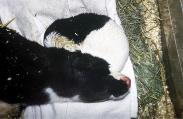
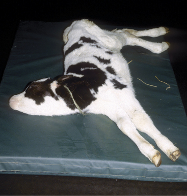
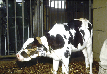
This is the most common brain malformation observed in the northeastern United States and is the result of an in utero infection with the bovine virus diarrhea virus (BVDV) agent, usually between 100 and 200 days of gestation. The inflammation peaks about 14 days after infection and resolves before birth. The small malformed cerebellum seen at birth reflects atrophy of the already differentiated cerebellar parenchyma at the time of the infection and hypoplasia from the destruction of the embryonic precursor cells primarily in the external germinal layer (Figure 12-1). In most affected calves, the cerebellum is largely absent with only a few remnants of cerebellar folia remaining (see video clips 25 to 27). Clinical signs vary. Some calves are unable to stand and often thrash around in their attempts to get up, and they exhibit periods of opisthotonos and sometimes abnormal nystagmus. Others can stand and walk but have a base wide posture, stagger, and weave from side to side with a hypermetric gait and balance loss (Figure 12-2).

Figure 12-1 Brain from a 3-week-old calf with cerebellar hypoplasia and atrophy caused by in utero BVD infection. Amazingly the calf was able to stand.

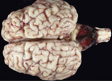
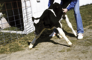
Figure 12-2 A 3-week-old calf with severe cerebellar hypoplasia and atrophy (see Figure 12-1) that was able to stand; notice the base-wide stance and lowered head position.
It is important to obtain a necropsy diagnosis for these calves because their clinical signs do not differ from a possible genetically induced cerebellar malformation. The latter has been observed in Angus and Scottish Highland calves with a symmetrically reduced cerebellar size but no gross or microscopic evidence of any destructive process. In addition, there is no trapezoid body on the ventral surface of the rostral medulla, but there is an abnormal band of parenchyma passing across the fourth ventricle just caudal to the cerebellar peduncles with a nucleus at each end. This may be the trapezoid body and the cochlear nuclei in an abnormal position that cannot be explained by an in utero viral infection. The fourth ventricle is remarkably reduced in size.
Congenital Cerebellar Function Disorder
On a few occasions, we have seen Holstein calves unable to get up at birth that exhibit opisthotonos and extensor rigidity on attempts to rise. If assisted, voluntary movements are delayed and overreactive. These calves are unable to balance and have abnormal nystagmus but are alert, responsive, and visual. Their anatomical dysfunction is primarily cerebellar. At necropsy, there are no gross or microscopic lesions anywhere in the nervous system. This is presumed to be a functional cerebellar disorder that may be inherited, but the latter remains unproven.
Hydranencephaly
This is primarily a cerebral abnormality in which the neopallium is reduced to a thin transparent membrane of pia and glial tissue as a result of complete destruction of the cerebral parenchyma. The lateral ventricle, containing a huge volume of cerebrospinal fluid (CSF), expands to take up the space vacated by the parenchymal loss. This is compensatory hydrocephalus. The hippocampus and olfactory bulb and peduncle and the basal nuclei are usually spared. The skull has a normal shape because CSF circulation is not obstructed in these calves. Akabane or bluetongue virus in utero infection at around 125 days of gestation is a recognized cause of this lesion. BVDV has also been reported as a cause, but this has not been seen in the northeastern United States where the BVDV-induced cerebellar lesion is common. The Aino and Chuzan viruses have also been implicated. The lesion is probably the result of the destruction of mitotically active progenitor germinal cells, as well as a vasculitis of the branches of the arterial circle that compromises the blood supply to the developing cerebrum. If the lesion is limited to cerebral hydranencephaly, the clinical signs are prosencephalic, and the animal will be able to ambulate but will be obtunded and blind.
Congenital Obstructive Hydrocephalus (Hypertensive)
Inherited forms are described in numerous breeds: Hereford, Charolais, Dexter, Ayrshires, and Holstein. The obstructive hydrocephalus is often accompanied by other brain malformations, which will influence the character of the clinical signs. A common cause of the obstruction is a failure of the mesencephalic aqueduct to develop normally. The latter may be associated with the presence of a single structure representing the rostral colliculi. The cause of this mesencephalic malformation is unknown in cattle but is inherited in laboratory rodents. Clinical signs will be prosencephalic, but brainstem and cerebellar signs may be present if there is significantly increased intracranial pressure.
Meningoencephalocele
This malformation occurs along the midline of the calvaria through an opening referred to as cranioschisis or cranium bifidum. The size of the extracranial accumulation of CSF may be extensive, producing a soft fluctuant pendular skin-covered structure (Figure 12-3). Although it is possible that some of these malformations may just be meningoceles, microscopic study of the tissues containing the CSF usually reveals a thin layer of brain parenchyma associated with the meninges beneath the skin, and therefore these are meningoencephaloceles.
Lipomeningocele
These also can occur along the midline of the calvaria or vertebral column through a cranioschisis or spina bifida, respectively. They consist of fat-filled meningeal tissue continuous with the falx cerebri in the head or the dural surface in the vertebral canal. With no associated neural tube malformation, there are no neurologic signs in these animals. The cause is unknown.
Complex Nervous System Malformation
A unique multifocal bone and neural tube malformation described in calves has been called an Arnold-Chiari malformation, presumably because of an assumed similarity to a human malformation given this eponym. Although there are some similarities, the distinct differences in the bovine disorder make use of this eponym incorrect. These calves are usually born recumbent and unable to coordinate their limb and trunk function to stand. They often exhibit opisthotonos and abnormal nystagmus. There is a sacrocaudal spina bifida with a meningomyelocele, a malformed tail, and associated loss of tone and reflexes in the anus and tail. At necropsy, the meningomyelocele consists of sacrocaudal nerves connecting from their spinal cord segments in the exposed vertebral canal into the skin-covered swelling over the spina bifida. The ganglia for these nerves are located in this skin. Myelodysplasia is present in the sacrocaudal segments. In the head, the cerebellum is flattened and elongated into a cone-shaped structure, and it is displaced into the foramen of the atlas and cranial axis along with the medulla. The associated CNs are elongated to extend back into the cranial cavity to exit through their respective foramina. There is a bilateral abnormal extension of each occipital lobe into the caudal cranial fossa space vacated by the cerebellum. These abnormal extensions of the otherwise normal occipital lobes pass ventral to the tentorium, which results in a groove on the lateral side of each of these extensions. These are not herniations of the normal occipital lobes. This malformation has been sporadically recognized in calves since the early 1900s.
Partial Diprosopus/Dicephalus
Occasionally calves are born with partial duplication of the face (diprosopus). This usually consists of varying degrees of two separate nasal regions; therefore four nares, parts of two lower jaws, and three orbits with the central one enlarged to accommodate two separate or fused eyeballs. The cranial region is broad, but there are two normal ears and a single normal atlantooccipital joint. These calves have four cerebral hemispheres (one for each naris formed from the embryonic olfactory placode, which gave rise to the olfactory nerves). Each cerebral hemisphere has a normal olfactory bulb, which resides in the cribriform plate related to each nasal cavity. There are four ethmoid bones. Two diencephalons are present (one for each set of eyes, two pairs of optic nerves, and two optic chiasms). The brainstem usually becomes single somewhere in the mesencephalon. The pons, medulla, and cerebellum are single structures. This is a partial dicephalus. These calves are usually born alive but are recumbent and unable to stand.
Prosencephalic Hypoplasia-Telencephalic Aplasia
Calves with this sporadic unique malformation are alive at birth and unable to stand. Their cranium is flattened between two normal orbits with normal eyeballs. A dorsal midline skin defect is present at the level of the caudal aspect of the orbits. There usually is a slight bloody discharge from this opening, which probably contains CSF. The skin tissue surrounding the opening is continuous caudally with a malformed diencephalon at the rostral portion of the brainstem. There are no cerebral hemispheres, just a malformed brainstem and cerebellum. There are no recognizable geniculate nuclei, no mesencephalic colliculi, and the cerebellum is elongated. With the exception of the olfactory nerves, all the remaining CNs are present, including the optic nerves that extend to the two eyes. In humans, this defect is called anencephaly, which is inappropriate because there is a brainstem and cerebellum present. There is no adequate term for this combination of malformations, and we have chosen to call this prosencephalic hypoplasia with telencephalic aplasia. The cause is unknown in cattle but has been blamed on folic acid deficiency or hyperthermia in humans.
Congenital Tremors
Hypomyelinogenesis
Failure to develop normal central nervous system (CNS) myelin can be the result of an inherited defect in oligodendroglial function or an in utero infection of the fetus that interferes with this process. Some strains of the BVDV have been implicated. An inherited hypomyelinogenesis has been reported in Jersey calves. Calves are usually recumbent at birth, and any muscular activity elicits diffuse whole body tremors. The more excited the calf becomes and struggles to move, the worse the tremor. It disappears when the calf is completely relaxed. These calves are usually alert, responsive, and visual. Occasionally calves affected by an in utero BVDV infection will improve over a few weeks, suggesting they became able to develop CNS myelination (see video clips 28 to 30).
Axonopathy
We recently studied a group of related Holstein calves that at birth were usually able to stand and walk but had a constant coarse tremor primarily of the trunk and pelvic limbs. At necropsy, they all had a diffuse primary axonopathy throughout the spinal cord with secondary demyelination.
INFLAMMATION OF THE BRAIN
Meningitis
Etiology
Gram-negative septicemia in neonates is the most common cause of meningitis in dairy cattle. Calves given inadequate amounts of high quality colostrum have insufficient levels of passively acquired immunoglobulins to fend off opportunistic organisms. Septicemia may originate in umbilical infections or more commonly by oral inoculation of pathogens. Gram-negative organisms such as Escherichia coli, Klebsiella sp., and Salmonella sp. predominate, with E. coli being the most common organism to infect neonatal calves. In colostrum-deficient calves, Streptococcus spp. may also cause bacteremia with meningitis, endophthalmitis, and peritonitis. Although any opportunistic or environmental organism may infect a calf with inadequate amounts of passively acquired immunoglobulins, only extremely pathogenic organisms will cause meningitis in a calf having adequate immunoglobulin supplies.
Although meningitis is a sporadic disease on well-managed farms, endemic problems may develop when calf husbandry is poor. Certain strains of causative gram-negative or less commonly gram-positive bacteria seem to result in meningitis in a high percentage of calves that develop septicemia. The owner may report similar signs in other calves that have subsequently died. An unusual strain of E. coli was identified as the cause of endemic meningitis in a “baby beef” Holstein calf operation. Affected calves were 2 to 5 months of age. This outbreak represents the first time that we have seen E. coli meningitis in calves of this age that are immunocompetent.
Acute bacterial meningitis in adult dairy cattle is not as common, and most cases are sporadic. Confirmed sporadic cases of meningitis in adult cows have been caused by septicemic spread of bacterial organisms from acutely infected organs such as the mammary gland or uterus, or foci of chronic infection such as traumatic reticuloperitonitis abscesses. Coliform mastitis may be the most common predisposing cause of sporadic bacterial meningitis in adult cattle in our practice area. Mycotic encephalitis has been observed as a sequela to mycotic mastitis and mycotic rumenitis with subsequent embolic septicemia. Direct extension of chronic infections such as pituitary abscesses and chronic frontal sinusitis may also result in meningitis in adult dairy cattle. When multiple cases of acute meningitis occur within a herd of adult cattle, Histophilus (Haemophilus) somni infection should be suspected. It must be emphasized that H. somni may cause meningitis rather than thrombotic meningoencephalitis (TME) in adult dairy cattle. Rarely, TME is observed in dairy heifers between 6 and 24 months of age.
Signs
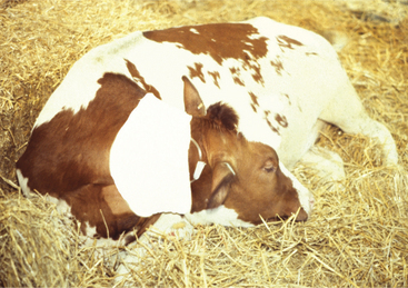
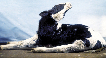
Signs of meningitis in neonatal calves may be overt, with classical fever, somnolence, intermittent seizures, head pressing, and blindness, or be masked by hypovolemic shock and collapse in overwhelming septicemias. When meningitis precedes other major organ infection, signs of fever, depression, head pressing or “headache” appearance, seizures, and cerebral blindness signal the diagnosis (Figure 12-4). The gait is stiff, and the head is often held straight, with the muzzle extended. The condition is painful, and the animal may appear to have a “headache” with the eyelids partially closed and the head and neck extended. However, when meningitis coexists with other organ infection such as uveitis, septic arthritis, and omphalophlebitis, it may be difficult to recognize specific signs of meningitis. Overwhelming septicemia results in rapid deterioration of the neonate such that shock may mask clinical signs of meningitis (see video clip 31). Some calves affected with meningitis have opisthotonos—perhaps caused by cerebral inflammation and edema exerting pressure on the cerebellum and caudal brainstem (Figure 12-5). Affected calves are generally between 2 and 14 days of age with the mean being 6 days of age.

Figure 12-4 Two calves with meningitis. The calf on the left is head pressing into the wall. The calf on the right is unaware of its surroundings and has hay in its mouth but is not chewing. Both calves are blind.

Figure 12-5 A 10-day-old Holstein calf with bacterial meningitis causing severe opisthotonos. The CSF was grossly abnormal.
Adult cattle affected with meningitis usually have fever and profound depression. A stiff, stilted gait and “headache” appearance (stargazing or continually pressing head or muzzle against an object) are common (Figure 12-6), but seizures are less common than in calves. Inflammation of the visual cortex can result in blindness with normal pupillary function.

Figure 12-6 An adult cow exhibiting depression with a rather rigid “stargazing” appearance. The cow had a fractured skull, bacterial sinusitis, and meningitis.
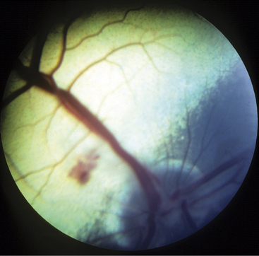
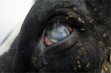
Meningitis caused by H. somni is acute, with affected cows becoming extremely depressed within a few hours. Fever usually is present and may be as high as 106.0° F/41.11° C. The depression progresses over 12 to 24 hours to total inappetence and somnolence, and the affected cow may be unable to rise (Figure 12-7). Depression is so severe that presence or absence of vision may be difficult to determine, and occasional seizures are observed in some patients. Affected cows die within 24 to 48 hours of onset unless treated specifically for H. somni. Herds experiencing H. somni meningitis often have multiple cases over a period of several months, until appropriate diagnostics and preventive measures are used. TME caused by H. somni, although rare, does occur in growing dairy heifers and causes acute severe neurologic disease that may be accompanied by retinal lesions (Figure 12-8).

Figure 12-7 A 13-month-old recumbent red and white Holstein with H. somni meningoencephalitis. The heifer was treated with ampicillin and supportive treatment and recovered in 1 month. This favorable outcome is unusual in such a severely affected animal.
Diagnosis
Clinical signs coupled with a CSF analysis confirm the diagnosis. Increased values for protein (normal 5 #40 mg/dl) and white blood cells (WBCs) (normal 5 #6 nucleated cells/ml) are present in the CSF, and the WBCs are mostly neutrophils in acute cases. In subacute cases, macrophages may predominate. The fluid can appear normal on visual examination, or it can be grossly discolored (red to orange). Neonatal calves showing neurologic signs that also have omphalophlebitis, uveitis, or septic arthritis should be suspected of having meningitis.
Bacterial cultures of the CSF and blood are indicated to determine the exact causative organism. Serum protein and immunoglobulin levels should be evaluated in neonatal calves to assess adequacy of passive transfer of immunoglobulins and thereby assess calf management procedures.
Diagnosis may be more difficult in adult cattle with meningitis secondary to acute or chronic infections elsewhere in the body. These cattle have been ill for variable lengths of time, and the developing signs of meningitis may be mistakenly assumed to be progressive systemic illness associated with failure to respond to therapy for the primary condition. Depression, an extended head and neck, head pressing, blindness with intact pupillary light responses, and seizures are all possible signs that may exist in individual patients. To repeat, CSF evaluation is necessary for diagnosis and will yield increased protein and WBCs—primarily neutrophils in acute cases and macrophages in more chronic cases.
Treatment
Broad-spectrum antibiotics constitute the primary treatment for meningitis in calves and adult cattle. Although the blood-brain barrier normally interferes with effective CSF levels for most antibiotics, the barrier is compromised by inflammation in meningitis patients. Therefore most antibiotics will enter the CSF in higher levels than would be possible in the healthy state. Antibiotics should be chosen based on the likely causative organism. For example, in neonatal calves, the anticipated cause would be a gram-negative organism such as E. coli, and appropriate antibiotics would include an aminoglycoside or other antibiotic effective against E. coli (e.g., ceftiofur 2 to 4 mg/kg twice daily or amikacin 20 mg/kg once daily plus ampicillin 10 mg/kg twice daily, or florfenicol 20 mg/kg twice daily). If amikacin is used, fluids should be given and proper meat withdrawal time advised. Although not permitted in North America, enrofloxacin would be an excellent antimicrobial selection for gram-negative meningitis. In adult cattle with secondary meningitis, the likely cause of the primary disease (e.g., mastitis, metritis) should be addressed when choosing a systemic antibiotic. Gram stain evaluation of CSF may be rewarding in some cases and thereby guide antibiotic selection. When H. somni is suspected, ampicillin (11.0 to 22.0 mg/kg twice daily) and florfenicol (20 mg/kg twice daily in replacement heifers) are reasonable antibiotic choices. Without early treatment, the prognosis for recovery is grave. Some calves that are aggressively treated too late with proper antibiotics may live for several days but never regain reasonable mentation and have necrotic lesions in the brain at necropsy.
Supportive treatment with a single dose of corticosteroids (5 to 10 mg of dexamethasone and/or mannitol 0.5 mg/kg slowly intravenously [IV]) may help decrease life-threatening inflammation and cerebral edema associated with meningitis. Some practitioners administer nonsteroidal antiinflammatory drugs (NSAIDs) instead of corticosteroids. If the inflammation cannot be immediately controlled, the calf will probably die despite proper antimicrobial therapy. Seizures may be controlled with 5 to 10 mg of diazepam in neonatal meningitis patients.
Prevention
Adequate passive transfer of immunoglobulins through well-managed colostrum feeding of each newborn calf is the most important method of prevention. Dipping navels and providing a clean, dry environment will minimize opportunities for navel infection, septicemia, and meningitis. Herd vaccination against H. somni is indicated whenever meningitis or TME is found to be caused by this organism.
Brain Abscesses and Pituitary Abscesses
Etiology
Brain abscesses, similar to abscesses affecting the spinal cord, usually arise from embolic spread of bacteria from distant sites of infection or during septicemic episodes. Calves develop brain abscesses most commonly from umbilical sepsis and extensions from otitis media/interna, whereas those in adult cattle have been associated with chronic infections, such as abscesses resulting from hardware disease, chronic musculoskeletal abscesses, or rumenitis. In addition, direct extension from chronic frontal sinusitis and bacterial seeding associated with nose rings in bulls are other potential causes of brain abscesses in adult cattle. Although the relationship with frontal sinusitis is obvious, the inferred higher risk of cattle or bulls with nose rings for brain or pituitary abscesses is very interesting. Theories to explain this phenomenon center around the complex rete mirabile circulation that encircles the pituitary region and is suspended in the cavernous sinuses, which drain the nasal cavity. Arcanobacterium pyogenes is the most common organism isolated from brain abscesses in cattle.
Signs
Signs vary tremendously, depending on neuroanatomic location of the brain abscess. Initial signs such as mild depression, dysphagia, hemiparesis, and hemianopsia may be subtle and will frequently go undetected by the owner. As the abscess enlarges, varying degrees of visual disturbance, paresis, ataxia, profound depression, and CN signs become apparent. Head pressing may be observed (see video clips 32 and 33). Calves tend to be affected between 2 and 8 months of age, thereby being past the typical age for neonatal meningitis. If the abscess becomes sufficiently large, it will interfere with venous return of blood from the orbital region and cause exophthalmos (Figure 12-9). Adult cattle can be of any age. Depression and a stargazing attitude have been observed in cattle with cerebral abscesses. Bradycardia coupled with depression and a stargazing attitude has been described to indicate a pituitary abscess (Fox FH, personal communication, 1985, Ithaca, NY), but other signs such as blindness, dysphagia, or CN signs are possible. A review of pituitary abscesses found that approximately 50% had bradycardia in addition to other neurologic signs. The bradycardia may result from involvement of hypothalamus or may be caused by the anorexia.

Figure 12-9 A heifer with exophthalmos, swollen conjunctiva, salivation, and depression caused by a brain abscess. Cerebrospinal fluid was normal.
Abscesses localized to one cerebral hemisphere usually cause blindness with intact pupillary function in the contralateral eye (hemianopsia) as a result of optic radiation or cerebral cortical injury (Figures 12-10 and 12-11). Similarly, contralateral abnormal postural reactions would be anticipated with a normal gait in animals light enough to be hopped or a scuffing of the limbs when walked in a tight circle or over rough ground. Propulsive tendencies may appear also with large cerebral abscesses. Anorexia secondary to severe depression may be accentuated by specific CN dysfunction if the abscess directly or indirectly damages the brainstem. Some affected cattle continue to eat despite extensive space-occupying abscesses.

Figure 12-10 Calf with a brain abscess. The calf is profoundly depressed, unaware of its surroundings, has a “stargazing” head carriage with the head and neck turned to the right (pleurothotonos), and has right side hemianopsia and right hopping deficits. A left cerebral abscess was identified at necropsy.

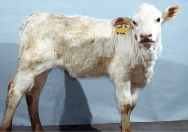
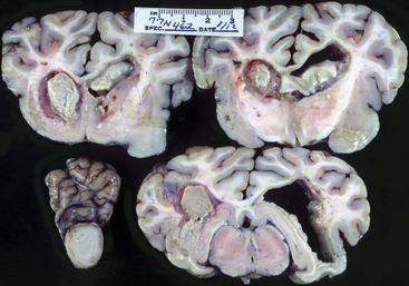
Figure 12-11 Sections of brain from calf shown in Figure 12-10. The left side of the photo illustrates the left side of the brain.
Diagnosis
Antemortem confirmation of brain or pituitary abscesses may be difficult. The neurologic signs are the most helpful to diagnosis—especially in young animals in which inflammatory lesions are more common than other intracranial disorders. Serum globulin should be assessed because it frequently is elevated in adult cattle with brain abscesses but may be variable in calves and young cattle. A neutrophilic leukocytosis may be anticipated, but in fact the hemogram often is normal.
CSF may or may not be helpful. CSF may be normal in early cases but will be profoundly abnormal in advanced cases of abscessation with both protein and WBCs elevated. Generally a high percentage, but not all, of the WBCs are mononuclear because of macrophage activity instigated by the chronic infection. Erosion of the abscess to cause leptomeningitis incites a neutrophilic pleocytosis in the CSF.
Radiographs of the skull occasionally show fluid lines consistent with gas-fluid interfaces in large, advanced, cerebral abscesses. CT and MR imaging procedures are the most reliable but are expensive and require general anesthesia.
Treatment
Other than long-term antibiotic therapy and potential drainage, therapy is limited and prognosis grave. We are unaware of successful surgery for brain abscesses in cattle, although this is occasionally possible in some other species. Symptomatic therapy with antibiotics and antiinflammatories may cause a slight improvement in the animal’s neurologic signs but is short-lived, and death is inevitable for most cattle affected with brain abscesses.
Listeriosis
Etiology
Listeria monocytogenes, a small gram-positive rod that is ubiquitous in soil, vegetable matter, and fecal material from humans and animals, is the cause of the most common meningoencephalitis of adult cattle. Although this facultative intracellular organism occasionally causes septicemia in young calves and abortion in adult cows, it is best known for the neurologic infection of the brainstem that is labeled listeriosis or “circling disease” in adult cattle and other ruminants. Use gloves when examining these animals because humans are susceptible to this infectious agent.
L. monocytogenes type 4b has been the most common serotype to cause meningoencephalitis in cattle. The organism is present in chopped forages such as corn silage and haylage owing to the presence of both soil and vegetable matter in these feedstuffs. Proper ensiling, wherein fermentation lowers the pH of the silage to, 5.0, kills or prevents multiplication of L. monocytogenes. However, improper ensiling as a result of excess dryness of the forage, lack of fermentation caused by trench ensiling, silage inoculants, and other variables may prevent the silage from achieving a pH of, 5.0, thereby allowing proliferation of L. monocytogenes. Corn silage is most incriminated as the forage source of organisms.
Infection is thought to occur following injury to mucous membranes of the oral cavity, nasal cavity, or conjunctiva with subsequent retrograde passage of the organisms via the sensory branches of the fifth CN (CN-V) to the brainstem. A possibility exists that cattle could become infected through the gastrointestinal tract with hematogenous spread to the brainstem as may occur in rodents and humans, but this route is thought less likely than following the peripheral nerve branches of CN-V.
Once established in the brainstem, the organism proliferates in the pons and medulla regions and may spread elsewhere. The trigeminal nerve and its neighboring CN nuclei are subject to injury as a result of neuritis, encephalitis, and meningitis. The classical histologic lesions of listeriosis consist of microabscesses subsequent to focal necrosis with abundant neutrophils and perivascular cuffing with mononuclear cells.
Fortunately, and rather inexplicably, given the common exposure of the whole herd to similar feedstuffs, the disease tends to be sporadic with only one animal in the herd affected. Endemics have been observed when two to six cattle become infected over a period of a few months, but this is much rarer in cattle than in sheep—where high flock morbidity is common. Calves are seldom affected, and the disease is seldom confirmed in cattle less than 12 months of age. This most likely coincides with less relative risk of exposure to feedstuffs containing L. monocytogenes in young animals but may be affected by increased dental eruption and therefore mucosal injury in young adult cattle.
Signs
Fever may be present, especially during the first few days of illness. The fever is not high (103.0 to 105.0° F/39.4 to 40.5° C), and absence of fever does not rule out listeriosis.
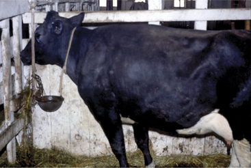
Depression coupled with a variable array of CN signs compose the major clinical signs of listeriosis in cattle. Classically the disease was known as circling disease because of the frequency of this clinical sign. The anatomic basis for this is unclear. Asymmetric involvement of vestibular nuclei with loss of balance and circling to that side is one explanation. However, the propulsive tendency to circle suggests involvement of extrapyramidal system nuclei such as the substantia nigra or the descending reticular formation. Although propulsion is a common prosencephalic sign, this portion of the brain is much less affected in listeriosis. Patients may circle until they collapse from exhaustion or eventually wander into solid objects. Stanchioned cattle constantly push or propel themselves into the stanchion in an effort to circle (Figure 12-12).

Figure 12-12 This cow with listeriosis would circle constantly to the left until finding respite by securing her muzzle between the water pipe and wall of the box stall.
Anorexia, or perhaps an inability to eat, is present in most cattle affected with listeriosis and may be caused by specific CN deficits in CN-V, -VII, -IX, -X, and -XII, as well as depression. Inability to drink frequently accompanies the inability to eat but is not present in all cases. Individual or combinations of CN injuries unique to each patient may occur (Figure 12-13) (see video clips 34 and 35).

Figure 12-13 Listeriosis patient having multiple cranial nerve deficits including left side CN-VII and CN-VIII, as well as CN-V, -IX, -X, and -XII. The right ear is drooped here.
General signs of depression characterize brainstem disease but may be accentuated by dehydration and acid-base deficits in cattle affected with listeriosis.
Lesions of CN-V motor nucleus or mandibular nerve create weakness in the muscles of mastication. When severe and bilateral, a dropped jaw results (Figure 12-14). When mild, weakness may be appreciated during manual efforts to open the patient’s mouth. Although this lesion may be unilateral, it is only obvious clinically when bilateral. Difficulty in prehension and mastication of food results.

Figure 12-14 Listeriosis patient with major signs of depression and inability to close her jaw (CN-V) and swallow (CN-IX and -X). Tongue tone was normal.
Lesions of the abducent nucleus (CN-VI) are not common but, when present, cause a distinct medial strabismus.
Facial nerve deficits caused by lesions involving the facial nucleus or the intramedullary components of the facial nerve are a very common sign of listeriosis and often are unilateral, causing a drooped ear, ptosis, and flaccid lip (Figure 12-15). Very early cases or cases recovering from complete facial nerve paralysis occasionally have facial nerve irritability evidenced by eyelid or lip spasticity in response to noxious stimuli. Although unilateral deficits in CN-VII are classic for Listeria meningoencephalitis of cattle, the deficits may be subtle, incomplete, or bilateral and therefore require careful evaluation during the neurologic examination. Exposure keratitis is the major ophthalmic complication found in listeriosis patients and results from facial nerve dysfunction and subsequent failure of tear distribution to prevent corneal desiccation or injury. Additionally, involvement of the parasympathetic facial nucleus may cause a decrease in the aqueous phase of the tear secretion. Exposure keratitis can rapidly progress with resultant deep corneal ulceration, uveitis, corneal perforation, and endophthalmitis unless addressed promptly. Endogenous uveitis with hypopyon or endophthalmitis has been suggested as possible ophthalmic complications by some authors, but in our experience, exposure keratitis and exogenous infection of the eye are the most common ophthalmic complications of listeriosis in cattle (Figure 12-16).

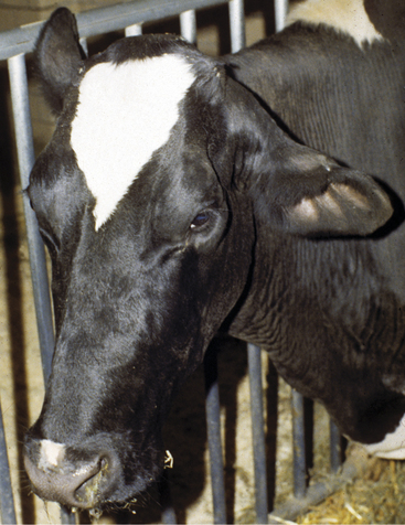
Figure 12-15 Listeriosis. Classical appearance of unilateral CN-VII paralysis with ear droop, ptosis, and flaccid lip.
Damage to vestibular nuclei affects central vestibular control, leading to head tilt, circling, and vestibular ataxia. Abnormal posture and truncal ataxia also are possible; when these signs are present, the cow’s trunk leans toward the affected side and is flexed so a concavity toward the affected side is present. When abnormal nystagmus is observed, the direction (fast phase) is variable, as expected with a central vestibular deficit. Adjacent unilateral lesions affecting reticulospinal UMN and spinocerebellar GP pathways may cause ipsilateral paresis and ataxia. Bilateral lesions in this area resulting in spastic tetraparesis and GP ataxia may be severe enough to cause recumbency.
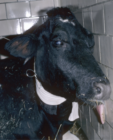
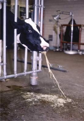
Lesions involving neuronal cell bodies of CN-IX and -X in the nucleus ambiguus cause dysphagia and salivation. Vomiting and/or bloat occasionally are observed (Figure 12-17) as an early sign of listeriosis in cattle and are thought to result from inflammatory irritation of the parasympathetic vagal neurons in the medulla.

Figure 12-17 An adult Holstein cow with an acute onset of depression, bloat, and vomiting. The cow had listeriosis and recovered with treatment.
Protrusion or weakness of the tongue is associated with lesions in the hypoglossal nuclei or the intramedullary components of CN-XII. Tongue protrusion caused by these lesions is accentuated if motor nuclei of CN-V lesions coexist, thereby allowing a dropped jaw.
Cerebellar signs also have been observed in listeriosis patients but are not common. Lesions caused by listeriosis are uncommon in the prosencephalon, and therefore blindness is very unusual. Spinal cord lesions are rare.
Diagnosis
Anorexia, depression, and possibly fever are the general signs that accompany specific CN signs in cattle having meningoencephalitis caused by L. monocytogenes. It is important to remember that “anorexia” may in fact be a result of inability to prehend or swallow food and water. A careful neurologic examination to confirm brainstem disease and specific CN deficits is essential when considering a diagnosis of listeriosis. In some cases, the deficits may only be detected on careful clinical examination.
In addition to the clinical signs, CSF analysis is the most valuable ancillary aid to support a diagnosis of listeriosis in cattle. With few exceptions, the CSF from listeriosis patients has elevated nucleated cells and protein levels. In one study 44 of 57 affected cattle had high leukocyte counts in the CSF. In addition, at least 50% of the nucleated cells are mononuclear cells, with macrophages being slightly more common than lymphocytes. CSF is usually obtained in the lumbosacral region unless the patient is recumbent and obtunded. The fluid is generally clear on visual inspection.
A complete blood count (CBC) may show mild leukocytosis and monocytosis, which is suggestive, but not absolute, of this disease. Unfortunately cattle affected with listeriosis do not frequently have the peripheral monocytosis that typically may be present in other species infected with this organism and that gave L. monocytogenes its name.
When salivation is obvious, an acid-base and electrolyte profile may be helpful for diagnosis and subsequent therapy because listeriosis patients can suffer profound salivary loss. Because the saliva of cattle is rich in buffer, patients so affected may have metabolic acidosis, low bicarbonate values, associated depression, and weakness.
Lactating cattle also may become ketotic because of continued (albeit reduced) milk production in the face of inappetence.
Many differential diagnoses exist for listeriosis, with rabies being the most important from a public health and medicolegal standpoint. Inner and middle ear infections may cause CN-VII and -VIII signs. Affected cattle are more alert and more able to eat and drink than are cows with listeriosis. In general, middle ear infections are common in calves up to yearling age, whereas listeriosis seldom occurs in cattle younger than 1 year of age.
Polioencephalomalacia (PEM), lead poisoning, and other diseases of the cerebral cortex can usually be differentiated from listeriosis unless the patient is recumbent or comatose, which limits the neurologic examination and a patient’s responses. PEM causes profound depression, bilateral cortical blindness with intact pupillary function, and may cause a dorsomedial strabismus in calves (not necessarily present in adult cattle). Opisthotonos may develop in advanced cases. Similarly, lead poisoning manifests with bilateral cortical blindness, depression, seizures, and bellowing, but no CN signs as seen with listeriosis. In addition, listeriosis does not result in blindness unless a severe exposure ulceration from facial nerve paralysis leads to uveitis or endophthalmitis in the ipsilateral eye. The lack of a menace response in listeriosis is a result of the facial paralysis, which also causes a lack of a palpebral reflex. Listeriosis rarely causes blindness.
TME or pure meningitis caused by H. somni can lead to acute signs of brain disease in young cattle. Although TME occurs mainly in beef cattle, we occasionally observe this problem in dairy heifers. Signs vary based on the multifocal nature of the septic thrombi within the brain, and CN signs are possible, as well as the typical cerebral signs and depression. Fever may be present in the acute phase, and blindness caused by chorioretinal hemorrhages and thrombosis is possible. The CSF, however, helps differentiate H. somni from listeriosis because, although protein values are elevated in both diseases, the nucleated cell count with H. somni usually is greatly elevated and consists primarily of neutrophils. The fluid may be grossly discolored on visual inspection.
Nervous ketosis can occasionally be confused with listeriosis in stanchioned cattle that become propulsive or constantly push forward into the stanchion, mimicking the propulsion seen in some listeriosis patients. However, a lack of CN signs, positive urinary ketones, history of early lactation, and, if necessary, a normal CSF would rule out listeriosis.
Subtle or mild cases of listeriosis have been confused with gastrointestinal disorders such as traumatic reticuloperitonitis. This can easily happen if the patient shows little or no evidence of CN dysfunction but cannot eat or drink. Subsequent dehydration or lack of water intake causes the rumen ingesta to become very firm and dry. Deep ventral abdominal pressure exerted on the rumen may lead to apparent painful responses that erroneously lead one to suspect peritonitis. In addition, occasional listeriosis patients show vomiting as one of their initial signs before other CN signs become apparent. Vomiting is more commonly associated with highly acidic diets, indigestion, irritation of reflex centers from ingested hardware, and other gastrointestinal disorders. Once again, a careful neurologic examination is essential, and a CSF analysis should be considered.
Space-occupying lesions in the cerebrum or brainstem (e.g., lymphosarcoma) or parasitic migration can rarely be confused for listeriosis.
Unfortunately rabies is the most difficult disease to differentiate from listeriosis when dysphagia or other CN signs are present. Because rabies can result in virtually any neurologic sign, it must be considered in the differential diagnosis of listeriosis in endemic areas. In general, CSF in rabies patients has fewer nucleated cells and protein than those found in listeriosis patients. In addition, the high percentage of mononuclear cells to neutrophils found in listeriosis patients is not typical in rabies, in which case small lymphocytes predominate. Because overlap may occur in these values, extreme caution is warranted for handling patients in rabies-endemic areas.
Treatment
Treatment usually consists of intensive antibiotic therapy with either penicillin or tetracycline, although other antibiotics are reported to be effective in vitro. Two major therapeutic obstacles exist to antibiotic therapy that dictate higher dosages than normally used for other susceptible bacteria:
Therefore we have chosen to use penicillin at 44,000 U/kg twice daily, either intramuscularly (IM) or subcutaneously (SQ), to treat listeriosis patients. This is used for at least 7 days before being reduced to either once daily or 22,000 U/kg twice daily, IM or SQ.
IV administered penicillin, 10 million units thrice daily, or ampicillin, 10 mg/kg thrice daily, would likely result in higher concentration in the CSF but is more expensive. Some clinicians prefer to use oxytetracycline HCl at 10 mg/kg twice daily, IV or SQ. Dosage reduction generally coincides with signs of obvious clinical improvement, such as a return to an ability to eat and drink. Although the exact duration of therapy will vary in each case based on severity of signs and many other factors, the treatment should continue for at least 1 week beyond apparent cure as based on appetite, attitude, and other factors. Most affected cows require 7 to 21 days of therapy. Premature reduction in dosage or discontinuation of treatment risks relapse in listeriosis patients. Some degree of facial neuronal signs may persist for a long time in recovered listeriosis patients, and neurologic signs tend to resolve in the opposite order of their original appearance. There may be no improvement in clinical signs for 7 to 9 days in some animals.
Fluid and electrolyte status may be very important to the well-being of listeriosis patients that lose the ability to drink. Cattle that cannot drink but are not salivating can be given water and balanced electrolytes through a stomach tube. This improves patient hydration and also softens the firm rumen contents and encourages rumen activity. Patients that are salivating should be monitored for buffer loss and will require bicarbonate replacement therapy and fluids. These may be given IV—although this is a more expensive route—or orally with substantial water to correct dehydration. Replacement therapy will be necessary daily until salivary losses stop. The depression and weakness that occur with severe metabolic acidosis may be confused with progression of the disease or lack of response to therapy for listeriosis. Depending on degree of salivary loss, 4 to 16 oz of sodium bicarbonate may be required daily to compensate. Cattle with facial paralysis also require frequent treatments of the affected eye with topical ointments to prevent keratitis and corneal ulcers.
Nursing care, including a well-bedded box stall with good footing, is essential to survival of cattle affected with listeriosis. Assuming intensive antibiotic therapy, fluid therapy, and supportive care are given, prognosis is fair to good for cattle infected with listeriosis that are ambulatory when the diagnosis is made. The prognosis for cattle that are recumbent and unable to rise at the time of diagnosis is very poor.
L. monocytogenes is capable of infecting humans and thus causing meningoencephalitis. This is especially true in the very young, the very old, and the immunocompromised person. Therefore public health concerns exist. Listeriosis patients may shed L. monocytogenes in their milk, and this fact requires veterinarians to warn owners and caretakers against the consumption of raw milk. Milk from these cattle should be discarded. Even pasteurized milk subjected only to low-temperature pasteurization may contain the organism. Pregnant cattle with the neurologic form of listeriosis may abort during the duration of their disease. The cause of the abortion is generally septicemic spread of L. monocytogenes to the uterus. Therefore handling of the fetus, placenta, and so forth should be done carefully.
Rabies
Etiology
Rabies virus is transmitted to cattle and other warm-blooded animals by bites from infected vectors such as foxes, raccoons, skunks, bats, and vampire bats. Cats and dogs are more routinely vaccinated against the disease, but unvaccinated cats and dogs also present a risk to cattle, humans, and other species. The rabies virus is a member of the genus Lyssavirus within the Rhabdoviridae family and is uniformly fatal to infected animals. Therein lies the tremendous fear of infection that the word “rabies” holds for humans. Public health and medicolegal implications are obvious.
Although aerosol transmission occurs in nature, mainly in bats within their caves, people and animals have been infected through aerosols in laboratory settings. Ingestion of infected tissues also may occasionally result in infection of carnivores. However, the primary means of transmission of this neurotrophic virus is through bites from an infected animal that inoculates virus-laden saliva into the tissue of a noninfected animal.
The virus replicates at the site of inoculation in an animal recently bitten. It then travels in retrograde fashion within the axons of peripheral nerves to spinal ganglia to the spinal cord and eventually the brain. The virus is then shed into the salivary and nasal secretions of the infected animal through centrifugal distribution following CN axons to these secretory glands. Therefore the virus is concentrated in saliva and nasal secretions—making these fluids the most feared source of exposure to uninfected animals or people.
Incubation periods vary widely. Most experts agree that 1 week is the minimum, but the range varies from 1 to 3 weeks, 10 to 60 days, or 3 weeks to 3 months; all authors agree that rare instances exist where the incubation may be as long as 6 months. Infection through bites closer to the brain (i.e., face and neck) may lead to shorter incubation periods than distal limb bites.
Clinical Signs
Clinical signs of rabies in cattle, as well as other species, are variable and may include spinal cord signs, brainstem signs including CN signs, cerebral signs, apparent lameness, genitourinary signs, gastrointestinal signs, and mixtures thereof.
Because of the variation in clinical signs of rabies, veterinarians practicing in endemic areas are more cautious of cattle with overt neurologic signs. Several points are important generalities when discussing signs of rabies in cattle.
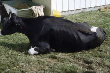
The clinical signs at the onset relate to the area of the body that is bitten and where the virus first enters the nervous system. Spinal cord signs are seen frequently. These may include subtle hind limb lameness or shifting of weight in the hind limbs that progresses to knuckling of one or both fetlocks (see video clip 36). Ataxia and weakness may follow these signs and progress until the cow needs help getting up or becomes completely paralyzed in the pelvic limbs (Figure 12-18). In some cases, there is a spastic uncontrolled flexion of the limbs. Associated with these lumbar and sacral signs, constipation, tenesmus, paraphimosis (males), dribbling of urine from bladder paralysis, and a flaccid tail and anus may become apparent. Therefore progressive signs of spinal cord or spinal nerve dysfunction should raise concern for rabies. With head bites, CN signs may occur initially.

Figure 12-18 A 4-year-old Holstein that was first noticed to be abnormal when she buckled on both hind limbs coming into the parlor. Within 2 hours, she was recumbent, would not eat, and began bellowing. Cerebrospinal fluid had a lymphocytic pleocytosis. She tested positive for rabies.
Cerebral signs include signs of progressive depression (“dumb form”) or aggression (“furious form”). Few veterinarians would fail to identify quickly any newly aggressive cattle as rabies suspects, but certainly nervous ketosis and hypomagnesemia would need to be ruled out. Other accentuated cerebral responses observed in rabies patients are hypersexuality (e.g., frequent mounting), localized or generalized pruritus that can progress to self-mutilation, seizures, tremors, alert eyes and ears despite paresis or ataxia, head pressing, bellowing, and opisthotonos. Blindness can occur but is not common.
Dysphagia, salivation, and a weak tongue are apparent in some cattle affected with rabies. An inability to drink usually accompanies these signs, which are reflective of pharyngeal paralysis. Bellowing is described as “peculiarly low pitched and hoarse and may progress to bubbly sounds prior to death.” Laryngeal paralysis associated with pharyngeal dysfunction may contribute to these sounds.
Affected cattle may have all or none of the signs described above. These are signs reported and observed in past cases, but no sign is pathognomonic for rabies.
The differential diagnosis is exhausting, but several common diseases should be considered. In the paralytic form with spinal cord signs predominating, sacral injuries from estrus activities and vertebral canal lymphosarcoma or abscesses should be differentiated from rabies. As discussed above, a personality change to furious or aggressive behavior should be differentiated from nervous ketosis, hypomagnesemia, or the occasional cow recently transported to a new location that simply “goes crazy” but has neither rabies nor nervous ketosis. With brain signs, the differential list is too long to consider simply because the brain signs possible with rabies are unlimited. In our experience, atypical listeriosis that causes dysphagia with or without tongue paralysis but without facial or vestibular nuclear signs is the disease most confused with rabies. However, in an advanced rabies case that is approaching coma, many encephalitic and toxic CNS diseases would need to be considered.
Diagnosis
CSF from rabies patients may be normal, have only elevated protein values, or have both elevated nucleated cells and protein. Most nucleated cells in the CSF of rabies patients are lymphocytes. No other premortem tests are helpful to the practicing veterinarian, and the brain from suspect animals must be submitted to the regional laboratory approved by the state health department for rabies testing. Currently fluorescent antibody (FA)-stained sections of brain offer the quickest and most accurate means of diagnosis. The FA test has replaced histologic examination of the brain for Negri bodies and the mouse inoculation tests. In addition, FA tests using monoclonal antibodies to epitopes of the virus can help distinguish the vector source of rabies (i.e., raccoon, fox, and bat) to aid epidemiologic studies.
No treatment is possible for rabies patients. However, cattle bitten by unknown assailants should have the wounds cleaned vigorously, as well as washed and disinfected, just as is done for people suffering bite wounds from animals of unknown rabies status.
When rabies is suspected in a cow or calf, gloves should be worn by the handlers and veterinarians during examination and treatment. A minimal number of people should be involved in treatment of the cow, and her milk should be discarded. If a cow is confirmed to have rabies, public health authorities should be consulted for advice on rabies prophylaxis therapy for any handlers that worked with the animal and had definite exposure to virus.
Rabies vaccination of cattle is now being practiced in many endemic areas and is a viable means to counteract the public and private anxiety regarding exposure to rabies while working with livestock. Vaccination also greatly reduces the likelihood of human exposure and subsequent expensive prophylaxis and treatment with globulin and human diploid vaccines. An entire herd (small size) of dairy cattle can be vaccinated for less than the cost of one human postexposure treatment. Therefore, vaccination of cattle in endemic areas is worthy of consideration. Veterinarians should be certain to use only vaccines approved for use in cattle because some modified vaccines are inappropriate for herbivores. At least two vaccines are currently available for use in cattle (RM Imrab3, Rhone Merieux Inc., Athens, GA and Rabguard TC, Smith Kline Beecham Animal Health, West Chester, PA). Both vaccines can be given initially at 3 months of age for primary immunization and repeated annually.
Pseudorabies (Aujeszky’s Disease, Mad Itch)
Etiology
This herpesvirus of swine is the cause of pseudorabies. Often a mild disease in swine, this disease is highly fatal in cattle and may cause signs similar to rabies—hence the name, pseudorabies. This is a rare disease in dairy cattle because pigs and dairy cows seldom are housed together. However, trends in agriculture change constantly, and diversification that includes swine and dairy cattle operations located on the same premises could occur, thereby risking spread of this virus from swine to cattle.
The virus is shed in the nasal secretions and pharyngeal secretions of infected pigs. Contact with cattle may include contamination of feedstuffs, contamination of wounds (because intradermal and SQ routes of infections are possible), and nose-to-nose contact. Infected brown rats also have been incriminated in carrying pseudorabies virus from farm to farm. Following infection, the incubation period is between 2 and 7 days.
Signs
Intense pruritus that may be localized or generalized develops, with licking, rubbing, and mutilation possible. This pattern has led to the name “mad itch” in cattle. However, many other neurologic signs are possible—similar to rabies—and fever usually is present. Peracute cases may die suddenly or have primary brain signs, which vary from salivation, pharyngeal/laryngeal dysfunction, dyspnea, bloat, ataxia, paresis, abnormal nystagmus, depression or aggression, and seizures. The course of the disease is 2 to 3 days, and although rare instances of survival have been noted in cattle, most infected cows succumb. Differential diagnosis would include rabies and many other neurologic diseases, but historical proximity of swine would be a key point.
Diagnosis
Serology, viral isolation from CNS (especially from a spinal cord segment supplying localized pruritus lesions), or edematous fluid from a localized lesion, and FA tests are possible. Tests continue to change and improve for this disease, and if this diagnosis is suspected, it would be best to contact a regional diagnostic laboratory for advice on sample collection. Rabies may need to be ruled out as well. No treatment exists. We are unaware of published CSF values for cattle affected with pseudorabies virus.
Bovine Herpesvirus Encephalitis
Etiology
Both bovine herpes virus (BHV) 1 and 5 may cause encephalitis in cattle. BHV1, a cause of abortion, infectious bovine rhinotracheitis, and pustular vulvovaginitis, only sporadically causes meningitis in cattle. On the other hand, BHV5 has marked neurotrophism, and in some parts of the world, particularly South America, it is a common cause of meningoencephalitis in cattle. BHV5 encephalitis can cause single disease on a farm or herd outbreaks, mostly in young replacement heifers, but the incidence in North America appears much lower than in South America. BHV5 meningoencephalitis may result from initial exposure to the virus or from a stress/corticosteroid reactivation at a later time. Clinical signs generally occur approximately 1 week after either initial exposure or reactivation of the virus. The virus invades the CNS via the olfactory mucosa following intranasal infection or reactivation. A trigeminal ganglionitis is found in infected calves, and this is an anatomical area of persistent infection in some calves.
Clinical Signs and Diagnosis
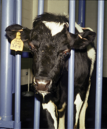
Clinical signs of respiratory disease may be concurrent with neurologic signs, especially with BHV1. Prosencephalic signs predominate and are usually accompanied by a fever. Most affected animals remain visual, which helps separate many of the infectious encephalitides from metabolic or toxic diseases affecting the cerebral cortex. I (TJD) have analyzed CSF on only one BHV encephalitic calf (Figure 12-19), and it had a lymphocytic pleocytosis. If BHV-infected cattle survive, they will seroconvert in 7 to 10 days. Animals that die with nonsuppurative encephalitis can be confirmed as having BHV by immunohistochemistry. Genomic analysis or polymerase chain reaction (PCR) can be used to differentiate between the two strains. The CNS lesion consists of a diffuse nonsuppurative meningoencephalomyelitis affecting both the gray and white matter.
Treatment and Prevention
Treatment is supportive and includes control of seizures when necessary, in addition to the use of NSAIDs. Corticosteroids would likely be contraindicated, although this is controversial. Although complete protection against either strain does not occur with vaccination, the modified live intranasal BHV1 vaccines have good efficacy against both strains.
Malignant Catarrhal Fever
Malignant catarrhal fever (MCF) is caused by a gamma herpes virus and sporadically causes fatal meningoencephalomyelitis in cattle. The virus that causes MCF in cattle in North America and Europe is sheep associated (ovine herpes virus 2). Most cases of MCF in cattle occur when affected cattle have had contact with sheep that are actively shedding the virus (especially weaned lambs). There have been some cases of MCF in cattle where a direct contact with sheep did not occur. The infection causes a vasculitis and lymphoproliferative reaction in many organs, including the CNS. The incubation period may be several weeks or more in cattle. We have recently identified the disease in pigs in contact with sheep.
Clinical Signs
Clinical signs are most common in cattle 1 to 2 years of age, and sporadic cases are the norm, although outbreaks can occur. There are basically two clinical forms: the head and eye or the intestinal form. Cattle with the head and eye form have a high fever, corneal opacity, nasal discharge, enlarged lymph nodes, hematuria, and diffuse neurologic signs. Similar to infectious bovine rhinotracheitis (IBR) keratitis, the corneal lesions often start at the limbus and spread centrally. Recovery with this form of the disease is rare. In the intestinal form, fever and diarrhea are the predominant clinical signs. Outbreaks are more common, as is recovery, compared with the head and eye form. We have collected CSF on only a few MCF/head and eye form cases. They had a remarkable mononuclear pleocytosis, and the exact type of some of the mononuclear cells was difficult to determine.
Diagnosis and Treatment
Diagnosis is based on signalment, clinical signs, history of sheep exposure (may be in distant past), and ruling out other diseases that may cause similar clinical signs (e.g., IBR). Hematuria, lymphadenopathy, and finding bizarre-appearing mononuclear cells in the CSF should help distinguish between the two diseases. An antemortem diagnosis can be made by performing PCR on whole blood (ethylenediaminetetraacetic acid [EDTA] anticoagulated sample). Postmortem diagnosis can be made by performing PCR on tissues. Lesions consist of a primary immune-mediated vasculitis with secondary parenchymal degeneration. Treatment is symptomatic.
Thrombotic Meningoencephalitis
TME is caused by Histophilus somni, formerly known as Haemophilus somnus. This small coccobacillus attacks vascular endothelium, causing a septic vasculitis with thrombosis. The parenchymal lesions of ischemic and hemorrhagic infarction are secondary to the primary vascular lesions that can occur anywhere in the CNS. In addition, similar vascular lesions can occur in the lung, heart, skeletal muscle, and joints. Death may occur acutely without evidence of neurologic signs. Pyrexia is present in clinically ill patients. This disease is more common in feedlot cattle than in pastured or dairy animals. In New York State pulmonary signs and lesions are the most common manifestation of this disease. Diagnosis and treatments for TME are discussed on p. 510.
Bovine Spongiform Encephalopathy
Although the purpose of this book is not to be all encompassing as regards exotic diseases but to concentrate on common problems in dairy cattle, bovine spongiform encephalopathy (BSE) deserves brief mention because of the threat of introduction into the United States, the possibility that at least one case has occurred in the United States, and the ensuing public health concerns. The abbreviation BSE should not be confused with sporadic bovine encephalitis (SBE or Buss disease), which is a chlamydial infection primarily seen in young beef cattle in the western United States. This is not an inflammatory disease but is caused by an unusual infectious agent.
Etiology
BSE is considered to be a form of scrapie in cattle. Scrapie is a disease of sheep and goats that is one of a group of diseases referred to as the transmissible spongiform encephalopathies (TSE). These include BSE, chronic wasting disease of deer, mink encephalopathy, and Creutzfeldt-Jakob disease of humans. The cattle disease first emerged in Great Britain in 1986 following the feeding of concentrates produced from slaughtered scrapie-infected sheep. Most investigators consider the infectious agent to be an altered host protein referred to as a prion.
One pathogenetic theory holds that BSE was most likely initially caused by scrapie-infected sheep-origin meat and bone meal products as part of calf starter rations or adult cattle concentrates. Another theory is that TSE developed in cattle and was exacerbated by feeding the tissues from infected cattle to other cattle. Public health concerns have focused on the similar features of the causative agent of Creutzfeldt-Jakob disease and kuru in humans to the prion-type causative agents found in scrapie and BSE. Because cattle were thought to have acquired this agent through ingestion, fears were raised relative to human consumption of meat products. Because the United States obviously has scrapie and chronic wasting disease as an endemic problem in sheep and deer, respectively, the threat of BSE in cattle raised in the United States seems to exist. However, great differences in amounts of sheep byproducts fed to cattle, as well as differences in rendering procedures, currently make the risk of BSE in U.S. cattle small. Additionally, there may be a species barrier, although not impermeable, to TSE transmission. There is also believed to be a genetic predisposition to the disease, and development of tests to determine resistant cows is being evaluated.
In addition to horizontal transmission, presumably from feeding contaminated ruminant tissue, there is vertical transmission of the TSE agent as well. BSE-infected cattle were three times more likely to have infected offspring than noninfected cows during the England outbreak. After banning the feeding of ruminant meat and bone meal to cattle in the United Kingdom, there has been a dramatic decrease in the incidence of BSE. In contrast, incidences in some other European and non-European countries have increased during the same period.
Signs
BSE occurs in adult dairy cattle 3 to 6 years of age or older. The disease is usually slowly progressive over 2 or more weeks. Hyperexcitability, an anxious expression, hypermetric ataxia, and hyperesthesia characterize the clinical signs. Affected cattle have facial and ear twitching, may kick repeatedly, and develop progressive ataxia and paresis that lead to stumbling, falling, and eventually recumbency. Only a small number of affected cattle display abnormal aggression (mad cow). Loss of weight is a significant sign in these cattle.
Diagnosis
Lesions consist of vacuoles in neuronal cell bodies or their processes in the neuropil sometimes associated with a mild gliosis but no inflammation. There is no serum antibody production, and the CSF is normal.
Immunodetection tests are available for detecting the prion in brainstem tissue. Currently in the United States, the disease has been confirmed in only a very few cows, which certainly justifies the many surveillance efforts that have been put into place.
DEGENERATIONS: METABOLIC AND TOXIC BRAIN DISEASES
Polioencephalomalacia
PEM describes a degenerative lesion of the gray matter of the cerebral cortex, a cerebrocortical necrosis for which there are many causes. These include thiamine deficiency, sulfur toxicity, lead poisoning, osmolality aberrations associated with salt and water imbalances, and hypoxia. Despite this, most clinicians equate PEM with thiamine deficiency, which is how it will be used in the following description.
Etiology
PEM or cerebrocortical necrosis is a thiamine-responsive disease that occurs in calves and adult cattle. In dairy calves, the disease usually is sporadic, but in grouped yearling heifers, the morbidity may reach 10% to 25%, similar to herd outbreaks in beef feeder calves or yearlings. The most common age for sporadic PEM in dairy calves is 2 to 8 months of age; calves less than 3 weeks of age are seldom at risk for PEM. Adult dairy cattle are only rarely affected.
The cause of PEM has been the subject of much research regarding thiamine metabolism, thiaminase activity in the rumen, the effect of various feedstuffs on rumen microbial flora related to thiamine production or destruction, and chemicals that alter thiamine levels in ruminants. Thiamine must be present in adequate levels to allow production of the coenzyme thiamine diphosphate, which is the active form of thiamine and helps to form red blood cell transketolase, and pyruvate and alpha-ketoglutarate dehydrogenases. This transketolase is important to the pentose phosphate shunt pathway for glucose metabolism in the bovine brain. Thiamine also participates as a cofactor in the Krebs cycle production of ATP. The brain is at great risk when thiamine is inadequate because of the brain’s dependence on aerobic metabolism where thiamine is a critical participant as a coenzyme. Within the CNS, specific groups of neurons are more susceptible to this interference with aerobic metabolism than others (i.e., neocortex, lateral geniculate nucleus, and caudal colliculus). It is important to appreciate that in severe cases the clinical signs represent a much more diffuse neuronal dysfunction than the distribution of histologic lesions seen at necropsy. This is a metabolic disorder that can disrupt neuronal function before causing ischemic-type degeneration or necrosis that is visible with the light microscope. The clinician’s objective is to stop this process as soon as possible before the neuronal changes become permanent.
Cattle normally produce thiamine as a result of rumen microbial activity. However, thiamine deficiency or alteration of normal thiamine production can be induced by a variety of means. High grain/low fiber diets are one of the most commonly encountered problems associated with field outbreaks of PEM. Such diets may alter the rumen flora to allow production of thiaminase type 2 by various anaerobes or other organisms. Similarly feedstuffs that contain thiaminase activity, such as bracken fern ribozyme and horsetail, may alter thiamine levels via thiaminase type 1. Type 2 thiaminase appears to be most important in causing PEM in cattle. Worming with levamisole or thiabendazole and tranquilization with acepromazine have also been incriminated by field experience. Although the feeding of amprolium, a thiamine analogue, has been associated with PEM, experimental cases were fed extremely high levels to reproduce the disease.
The exact etiology in sporadic cases usually is impossible to determine, although herd outbreaks warrant close analysis of possible contributing factors—especially concentrate versus roughage ratios.
Clinical Signs
Cerebrocortical signs predominate in PEM. Depression and anorexia are present in both calves and adults, but these signs may only be present for a short time before more overt signs of cortical disease become apparent. Blindness with intact pupillary function is one of the first signs observed because of the sensitivity of the visual cortex to the ongoing pathology (see video clips 37 and 38). Pupillary response to light may be lost in some recumbent cases as the disease progresses and presumably the oculomotor nerve is compressed. Head pressing and odontoprisis may be observed or an extended head and neck typical of cattle with “headache”-type pain. A slow shuffling gait is usually apparent if the animal is able to walk. Vocalization, as observed in some early cases in goats, is usually not observed in calves or adult cattle with PEM. A dorsomedial strabismus thought to be caused by involvement of the CN-IV nucleus is common in calves and yearlings but observed less often in adult cattle. Muscle tremors and salivation also may occur.
If the disease has progressed enough to cause recumbency, opisthotonos frequently is observed, and abnormal nystagmus, seizures, or coma is likely to follow (Figure 12-20). Untreated cases may die within 24 to 96 hours, depending on the severity of the metabolic dysfunction. CN signs other than possible dorsomedial strabismus usually are not present (Figure 12-21). Although optic disc edema has been reported to occur, we have never observed this in any calf or cow affected with PEM. Therefore in our opinion, blindness is entirely of cortical origin (Figure 12-22). Associated with either cortical blindness or cerebral edema, cattle may have a stargazing appearance (Figure 12-23).

Stay updated, free articles. Join our Telegram channel

Full access? Get Clinical Tree

