CHAPTER | 14 Neoplastic and Nonneoplastic Tumors


Intracutaneous Cornifying Epithelioma (keratoacanthoma, infundibular keratinizing acanthoma)
Diagnosis
Treatment and Prognosis
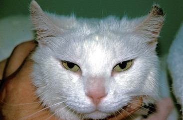
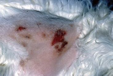
Feline Solar Dermatosis
Diagnosis
Treatment and Prognosis
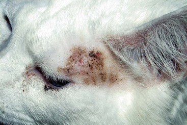
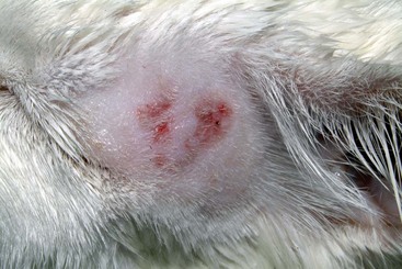
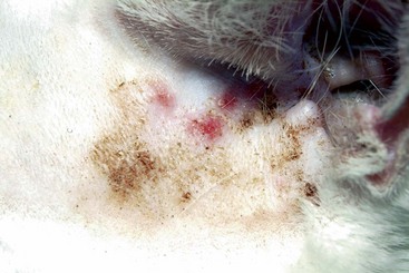
FIGURE 14-4 Feline Solar Dermatosis.
Same cat as in Figure 14-3. The crusts have been removed, revealing the erythematous papular lesions.

Canine Solar Dermatosis
Diagnosis
Treatment and Prognosis
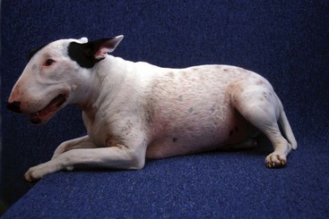
FIGURE 14-9 Canine Solar Dermatosis.
Generalized alopecia and erythema covering the face and trunk of a Bull terrier.
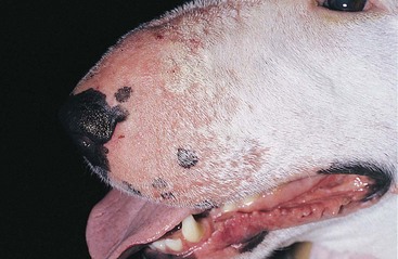
FIGURE 14-10 Canine Solar Dermatosis.
Alopecia and erythema with papular dermatitis on the muzzle.
(Courtesy D. Angarano.)
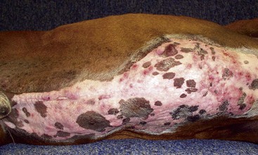
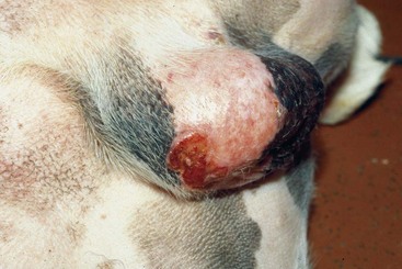
FIGURE 14-14 Canine Solar Dermatosis.
This focal area of ulceration on the scrotum of a Boxer had progressed to squamous cell carcinoma.
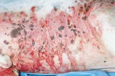
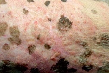
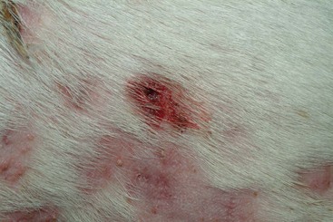
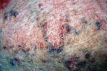
FIGURE 14-18 Canine Solar Dermatosis.
Severe erythematous dermatitis with a coalescing papular rash caused by sun exposure.
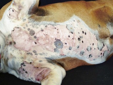
FIGURE 14-19 Canine Solar Dermatosis.
Severe erythema affecting only the nonpigmented areas of skin on the ventrum of a Boxer.
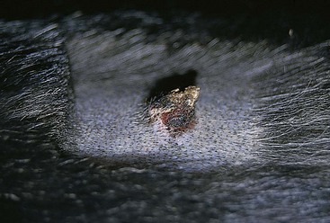
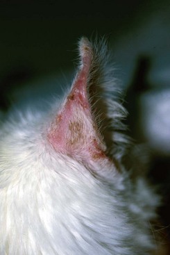
Squamous Cell Carcinoma
Diagnosis and Staging
Treatment and Prognosis
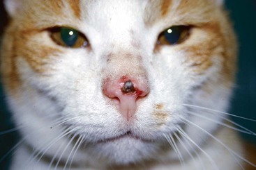
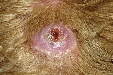
FIGURE 14-21 Squamous Cell Carcinoma.
A small, ulcerated tumor on the nonpigmented nasal planum of a cat.
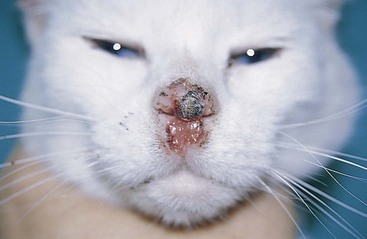
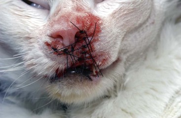
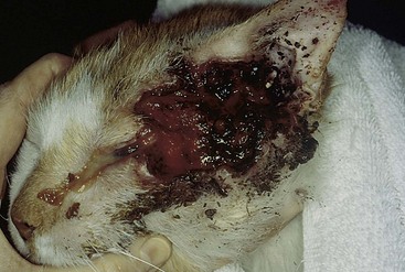
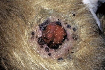
FIGURE 14-24 Squamous Cell Carcinoma.
Severe tissue destruction and tumor proliferation on the face and periocular tissue of a cat.
(Courtesy S. McLaughlin.)
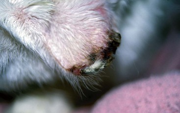
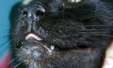
FIGURE 14-25 Squamous Cell Carcinoma.
Necrosis and crusting of the distal ear margin of an adult white cat.
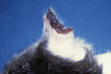
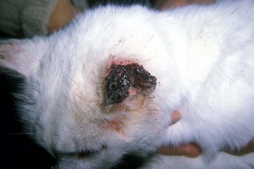
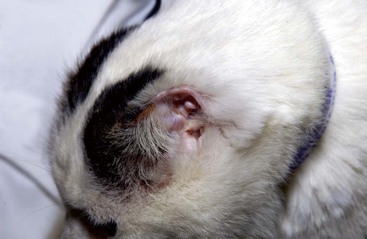
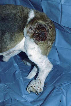
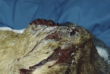
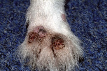
FIGURE 14-30 Squamous Cell Carcinoma.
Close-up of the dog in Figure 14-29. This raised tumor has a deep ulcer, with tissue destruction forming a central crater.
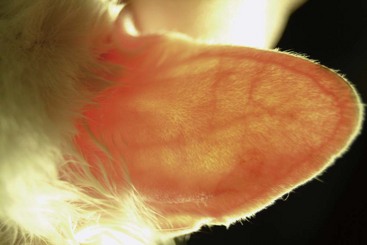
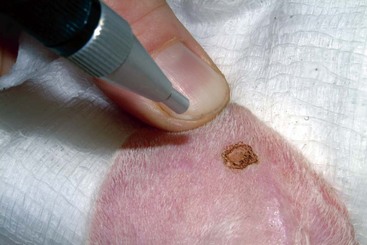
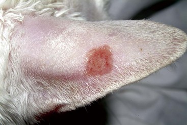
FIGURE 14-33 Squamous Cell Carcinoma.
Same cat as in Figure 14-32. The tumor has been ablated, leaving a focal area of ulceration.
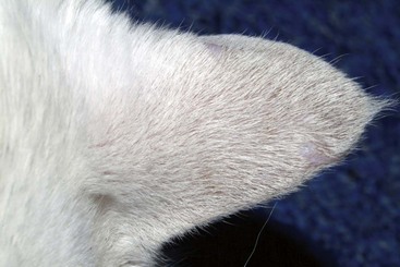
FIGURE 14-34 Squamous Cell Carcinoma.
Same cat as in Figure 14-32. Three weeks after treatment, the focal area has healed and the hair is regrowing. Early detection and therapeutic intervention provide better cosmetic outcomes.
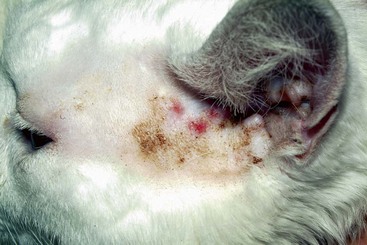
FIGURE 14-35 Squamous Cell Carcinoma.
Multifocal squamous cell carcinoma on the preauricular area of a white cat.
Bowen’s Disease/Multifocal Squamous Cell Carcinoma In Situ
Treatment and Prognosis
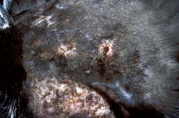
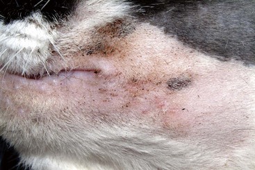
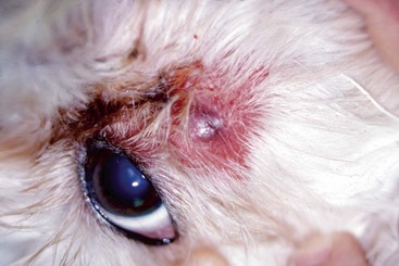
FIGURE 14-38 Bowen’s Disease.
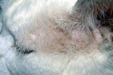
Multifocal, crusting, papular lesions on the face. Note the mild, subtle nature of the lesions.
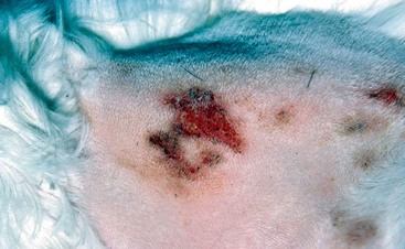
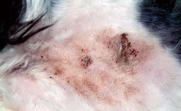
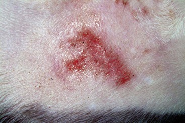
FIGURE 14-41 Bowen’s Disease.
Coalescing papules formed a plaque on the nonpigmented skin of this cat.
Basal Cell Tumor/Carcinoma
Diagnosis
Treatment and Prognosis
Hair Follicle Tumors
Features
Diagnosis
Treatment and Prognosis
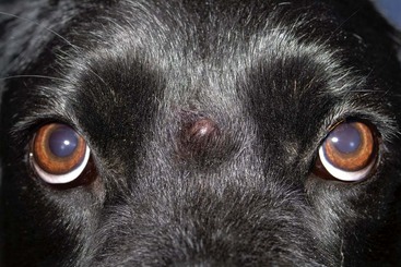
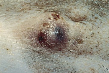
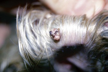
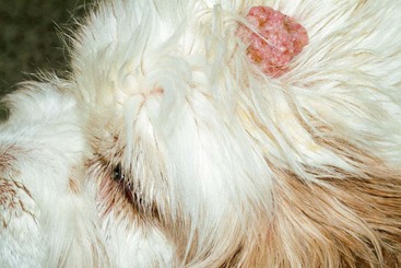
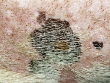
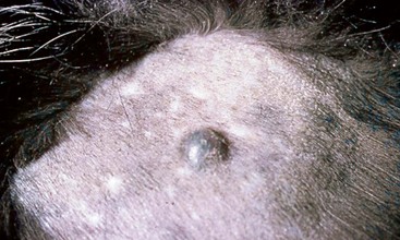
FIGURE 14-47 Hair Follicle Tumors.
A small, pigmented nodule. Note the similarity to basal cell tumors, apocrine tumors, and melanoma.
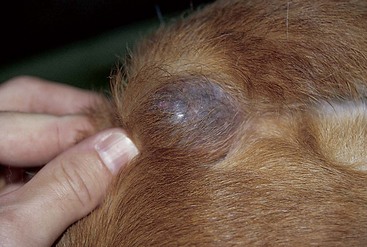
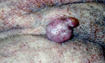
FIGURE 14-48 Hair Follicle Tumors.
This large cyst on the ventral thorax of an aged hound mix was associated with a follicular tumor.
Sebaceous Gland Tumors
Diagnosis
 Sebaceous hyperplasia/adenoma: cells exfoliate in groups and appear similar to normal sebaceous cells, with foamy pale blue cytoplasm and small dark nuclei.


Treatment and Prognosis
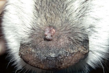
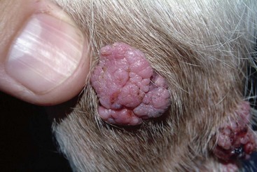
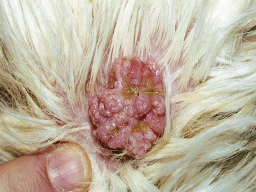
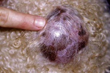
FIGURE 14-52 Sebaceous Gland Tumors.
This sebaceous adenoma on the nasal planum demonstrates the characteristic cauliflower appearance.
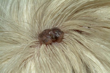
FIGURE 14-53 Sebaceous Gland Tumors.
This sebaceous adenoma had persisted for several years with little progression.