Neoplasia

Chapter 3 Neoplasia


The description of neoplastic disorders and incidence of neoplasia in farm animals has historically relied heavily on surveys conducted on animals at slaughter houses. The incidence of tumors reported in such surveys indicates that tumors are most common in cattle (0.23%) and are uncommon in sheep (0.002%), goats (0.009%), and pigs (0.004%).


Far fewer clinical descriptions of neoplasia in farm animals exist than do clinical reports of neoplastic diseases in dogs, cats, and horses. Reports of surgical treatment of tumors of farm animals are even less common. The changing face of livestock husbandry—with the increase in small “hobby” farms, sanctuaries, and petting zoos—will likely result in earlier detection of clinical signs associated with neoplastic disease and increased owner interest in pursuing surgery. With the advent of improved diagnostic procedures—in particular, ultrasound—detecting internal tumors at early stages is possible.


The following chapter describes neoplastic disorders of cattle, sheep, goats, and pigs that might present for surgical biopsy or surgical excision. As such, tumors of organs such as the heart and brain have been excluded. In many cases of farm animal neoplasia, extrapolation from information available on results of surgery on similar tumors in cats, dogs, and horses is the best that can be provided at this time. It is hoped that future editions of this or similar texts will be able to incorporate what should be a growing body of literature regarding surgical treatment of farm animal tumors.


Tumors of the Skin and Soft Tissue


Skin tumors are the most common neoplasms in farm animals. This is in large part caused by the high incidence of papillomavirus-induced lesions in the skin of cattle. Other tumors occur less frequently, and skin tumors in general are less common in sheep, goats, and pigs. Tumors of the soft tissue (subcutis and skeletal muscle) are uncommon and are included in this section because they are likely to present as mass lesions visible and palpable on external physical examination.


PAPILLOMA AND FIBROPAPILLOMA (PAPILLOMATOSIS)


Papillomas, commonly called warts, are the most common skin tumor of cattle. Shorthorn cattle appear to be predisposed to development of cutaneous papillomatosis. These tumors in cattle result from bovine papillomavirus infection and most often occur in animals less than 2 years of age. Viral infection of skin is thought to occur most often after trauma, infections, ectoparasites, or ultraviolet light exposure that damages the skin. Virally-induced cutaneous papillomatosis occurs less commonly in sheep and goats of any age, and a viral cause is suspected in affected goats. Papillomatosis is rare but can occur in pigs of all ages. Piglets can be born with congenital papillomas that may or may not be virally induced. Papillomas can also involve the eyes of cattle and the genitalia of cattle and pigs and are discussed in the sections on tumors of the eye and tumors of the female and of the male genital tract. Papillomas of the udder and teats in cattle, sheep, and goats are discussed in the section on tumors of the udder and mammary gland.


Bovine papillomas can become quite large and multinodular. They may be broad-based or pedunculated. They are alopecic and surface hyperkeratosis is typical. Exophytic growths are most common, although flat plaque-like growths are also possible. Lesions can occur anywhere on the body, but the head, neck and dewlap are common sites (Figure 3-1). In sheep, papillomatosis most often involves the skin of the face and legs. Goat papillomas most often occur on the head, neck, shoulders, and upper forelegs.
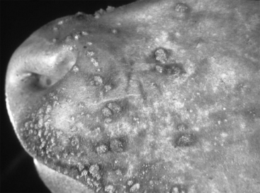


Raised lesions of the interdigital skin of adult cattle are a unique entity. These lesions have previously been called interdigital papillomas, but this disorder is now thought most likely to be induced by bacterial spirochetes rather than by papillomavirus. Papillomatous digital dermatitis is a more appropriate term, and these lesions are typically single, raised, painful growths often with surface finger-like projections of epithelium. Lameness, weight loss, and decreased milk production occur in cattle with interdigital papillomatous dermatitis. These growths can occur only in individual animals or can apparently spread to involve multiple animals.


Histologic evaluation of papillomas reveals two main types of proliferative lesions. Those with entirely squamous epithelial proliferations are classified as squamous papillomas, and those with proliferation of underlying fibrous connective tissue as well as of epithelium are classified as fibropapillomas. Papillomaviruses are known to be capable of inducing proliferation of both epithelial cells and fibroblasts. Identification of the spirochetes associated with bovine papillomatous digital dermatitis requires silver stains.


Papillomas in farm animals can be contagious, with an incubation period of about 2 to 6 months. Most papillomas that develop in young animals will spontaneously regress. Surgical excision of one tumor or use of immunostimulants have been said to speed the regression, but whether these procedures actually alter the natural course of the disease is not clear. Papillomas in adult sheep and goats less commonly regress spontaneously, and persistent tumors can undergo malignant transformation to squamous cell carcinoma (see Squamous Cell Carcinoma). Congenital papillomas in piglets can grow rapidly after birth. Surgical excision or cryotherapy of cutaneous and interdigital papillomas of cattle and of papillomas of sheep, goats, and piglets is often curative. In dairy cattle, treatment of interdigital papillomas with parenteral penicillin, ceftiofur, or topical oxytetracyline resulted in resolution of the lesions, which is good evidence that bacteria play an important pathogenic role in this disorder. Recurrence or subsequent development of new lesions, however, occurred in almost 50% of cases.


SQUAMOUS CELL CARCINOMA


Squamous cell carcinoma is the second most common skin tumor in almost all farm animals. Swine are the exception, as cutaneous squamous cell carcinoma is very rare in pigs. This is interesting, as the development of cutaneous squamous cell carcinoma has been linked to ultraviolet light exposure of thinly haired unpigmented skin, which most pigs have in abundance. Viral papillomas can progress to cutaneous squamous cell carcinoma, especially those on the udder (see Tumors of the Udder and Mammary Gland). Squamous cell carcinoma typically occurs in adult animals. In cattle, Herefords and Ayrshires are predisposed to squamous cell carcinoma, most likely because of their large areas of unpigmented skin. Aged ewes and Merino sheep are predisposed to development of squamous cell carcinoma, and both ultraviolet light exposure and exposure to photosensitizing plants are proposed etiologic factors. Saanen, Saanen cross, and Angora goats with white- or gray-haired areas appear to be predisposed to cutaneous squamous cell carcinoma. This tumor in goats has been reported to be more common in females, most likely because of the syndrome of udder papillomatosis and squamous cell carcinoma (see Tumors of the Udder).


Cutaneous squamous cell carcinoma causes a raised, proliferative, and ulcerated lesion and in cattle is most common at mucocutaneous junctions, such as the periocular skin and skin of the vulva. Squamous cell carcinoma in the skin of Merino sheep most often occurs on the ears and less commonly involves the muzzle, lower lip, and vulva. Those on the vulva occur primarily in sheep in which perineal surgery has been performed to reduce fly strike. Tumors in sheep are often multicentric. Squamous cell carcinoma of the ears of sheep can begin as a cutaneous horn, as a hyperkeratotic plaque, or can occur at the site of ear trauma such as from an identification punch. This tumor is rare in sheep less than 4 years of age. Squamous cell carcinoma in sheep can also occur in periocular skin. Cutaneous squamous cell carcinoma in goats is most common on the ears, udder, vulva, and perineum. Tumors in pigs can be single or multiple.


Histologic features are of well differentiated to poorly differentiated invasive squamous epithelium with associated sclerosis and inflammation. Atypical squamous cells indicative of neoplasia can be seen on cytologic preparations.


Metastasis of cutaneous squamous cell carcinoma can occur, most commonly to local lymph nodes. Metastasis is, however, typically a late event, preceded by a long period of local invasion. Immunotherapy of ovine squamous cell carcinoma has been shown to actually increase the rate of metastasis. Wide surgical excision, cryotherapy, hyperthermia, or radiation therapy of tumors before metastasis can be curative.


MELANOCYTIC TUMORS


Both benign and malignant melanocytic tumors occur in farm animals. Melanoma is most common in Sinclair miniature and Duroc-Jersey swine and is thought to be inherited in these breeds. Melanoma also occurs with some frequency in Hampshire and Iberian pigs. Melanoma does not occur in white swine and does not appear to be related to ultraviolet light exposure. Melanomas occur in the skin of cattle with some frequency, are relatively uncommon in goats, and are rare in sheep.


Melanomas in cattle occur most often in young animals. Tumors are either present at birth or develop within the first 2 years. Melanoma most commonly occurs in gray-haired cattle and in black-, or red-haired cattle, such as Angus. Melanoma in sheep occurs in the skin or the base of the horn in adult to aged animals, and Suffolk and Angora sheep are predisposed breeds. Melanoma in goats occurs in adult to aged animals. Angora goats may be predisposed, as is any goat of gray or brown coat color. Melanoma in predisposed swine breeds can develop at any age but are most often present at birth or develop within 1 year of age.


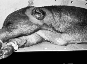
Melanoma is recognized clinically by areas of gray to black pigmentation within a solid fleshy raised mass. Melanomas occur within the dermis, the subcutis, or both. The overlying skin is often darkly pigmented, smooth, and partially to completely alopecic. Melanomas in cattle occur on the head (especially the jaw), neck, trunk, or legs. Most tumors arise in areas of pigmented hair. Size of tumors in cattle vary widely, from less than 5 cm to up to 25 cm. Melanoma in cattle most often occurs as a solitary lesion with intact overlying skin. Melanomas in sheep are often multiple and occur in the subcutis under areas of pigmented skin. Melanomas of goats occur most often in the perineum, with rare involvement of other sites such as the udder, coronary band, ear, and base of the horn. Melanomas in goats can be single or multicentric. Tumors of the perineum in goats vary from infiltrative to pedunculated and often are ulcerated. Affected goats often rub and lick the area, and secondary infection is common. Enlargement of local lymph nodes and poor body condition are also common in goats with melanoma. Melanomas in predisposed pigs can be either solitary or multiple, and occur as either flat plaquelike tumors or larger raised tumors, most often on the trunk (Figure 3-2).


Histologic and cytologic examination of samples from melanocytic tumors reveals the characteristic melanin-containing neoplastic cells, often admixed with heavily pigmented melanophages. Cellular pleomorphism and mitotic activity are variable. Those tumors with high mitotic indices are most often malignant. Evidence of local or epidermal invasion is also a good indicator of malignancy.


Melanocytic tumors that occur in young cattle are almost always benign and cured by wide surgical excision. Melanomas in older cattle can, however, develop metastases to local lymph nodes or internal organs. Melanomas of sheep and goats are most often malignant, with frequent metastases. Perineal melanomas of goats can exhibit widespread metastases to multiple lymph nodes; to bone; and to internal organs, including lungs, liver, adrenal glands and kidneys. Surgical excision of cutaneous melanomas in goats can be attempted, but death from metastatic tumors is common. The melanomas of predisposed breeds of pigs have an interesting behavior that has made them a useful animal model for the study of melanocytic tumors of people. The flat tumors typically spontaneous regress starting as early as 1 month of age, often leaving a halo of depigmented skin and hair. Larger, raised tumors often metastasize within the first year of life.


MAST CELL TUMORS


Mast cell tumors are most common in cattle and often involve the skin. Age of affected cattle ranges from 2 months to 12 years. Holstein and Holstein crosses may be predisposed. Cutaneous mastocytosis occurs in pigs 6 to 18 months of age. No reports were found of mast cell tumors in sheep or goats.


Mast cell tumors in the skin of cattle can be single or multiple. These tumors are raised, firm, tan nodular masses that can be ulcerated. Tumors can be discrete or infiltrative. Histologic and cytologic preparations are characterized by sheets of well differentiated to pleomorphic mast cells with admixed eosinophils. Mitoses are rare. Tumor necrosis, fibrosis and mineralization are common histologic findings in cattle. Porcine mastocytosis results in multiple nodular solid gray-white skin lesions that can be ulcerated. Tumors in pigs contain relatively homogeneous neoplastic mast cells admixed with eosinophils. Cutaneous mastocytosis in pigs may or may not have associated widespread visceral involvement.


Cutaneous mast cell tumors in cattle are associated with a relatively high rate of metastasis. Spread to lung and lymph node is most common, and metastasis to liver and muscle is also reported. Tumor cell morphology (i.e., well-differentiated vs. pleomorphic) does not appear to predict metastatic behavior, as even very well-differentiated mast cell tumors in cattle have undergone widespread metastasis. Wide surgical excision is the treatment of choice, and a guarded prognosis following surgery is warranted.


CUTANEOUS VASCULAR TUMORS


Cutaneous vascular tumors are most common in cattle but also occur in sheep, goats, and pigs. Tumors can be congenital or acquired. Congenital tumors may actually be hamartomas rather than true neoplasms.


Calves can be born with multiple cutaneous vascular lesions. Those that involve gingiva and tongue are described in Tumors of the Oral Cavity and Jaw. Cutaneous vascular lesions in neonatal calves are often described by owners as appearing to be “blood blisters.” Vascular lesions in neonatal calves can be disseminated throughout the skin and can also involve internal organs. In adult cattle, a syndrome known as bovine cutaneous angiomatosis occurs. Lesions can be single or multiple and often occur on the dorsum of the trunk. These tumors are soft, pink to reddish gray, can be sessile or pedunculated, and often bleed sporadically. Cutaneous vascular neoplasms occur in goats of all ages and are most often single, raised pink to red lesions that often bleed. In pigs, hemangiomas occur in the scrotal skin, especially of Yorkshire and Berkshire boars. Scrotal hemangiomas in pigs are often multicentric and progress from tiny purple papules to raised, hyperkeratotic lesions.


Cytologic evaluation of vascular tumors most often reveals only blood. Histologic evaluation is necessary to identify the proliferative endothelial cells lining vascular channels in these lesions. These channels vary from capillary (most common in congenital neoplasms of calves) to cavernous, and can contain a mixture of vascular structures. Very well differentiated lesions may be classified as hamartomas. Abnormal vascular channels lined by well-differentiated endothelium are hemangiomas, whereas tumors with cellular pleomorphism and mitotic activity are hemangiosarcomas.


Vascular lesions of all types can exhibit local invasion and recurrence after incomplete excision. Wide surgical excision of tumors, when possible, is often curative, as even cutaneous hemangiosarcomas rarely metastasize.


CYSTS


Cysts of the skin are uncommon lesions of cattle, sheep, goats and swine. They can be single or multiple, and can be congenital or acquired. Epidermal cysts (epidermal inclusion cysts), dermoid cysts, and follicular cysts are most common. Cysts are most common in Merino and Suffolk sheep, in which a hereditary basis is suspected. Cysts within the udder skin of older ewes involve mammary glandular epithelium and contain milk. Epidermal and dermoid cysts are less common in cattle. Cysts within the wattle of Nubian and Nubian cross goats are developmental anomalies present at or soon after birth that are suspected to arise from branchial cleft remnants. A hereditary basis for wattle cysts in goats is suspected.


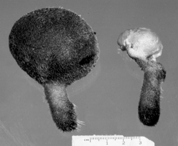
Cystic skin lesions in farm animals are most often asymptomatic, although their presence will adversely affect hide quality. Cysts can rupture and develop secondary inflammation with ulceration of overlying skin or can be discrete and nodular with normal overlying skin. Wattle cysts in goats are soft and fluctuant with normal overlying skin (Figure 3-3). Cystic skin lesions in sheep have been associated with development of carcinoma and with systemic illness.


Cytologic evaluation of epidermal, dermoid, and follicular cyst contents will reveal keratin, often admixed with cholesterol clefts and macrophages. Milk can be aspirated from udder cysts in sheep. Wattle cysts contain clear liquid. Surgical excision and histopathologic evaluation to identify the type of cells and structures within the cyst wall will allow for classification of the type of cyst present. Epidermal cysts are lined by keratinizing epithelium lacking adnexal structures. Dermoid cysts are lined by keratinizing epithelium with associated adnexa. Follicular cysts consist of dilated and keratin-filled hair follicles. Wattle cysts are lined by a single to double layer of cuboidal to columnar epithelial cells. When either a single or a small number of cysts are present, surgical excision is curative. Excision of udder cysts of sheep is neither necessary nor recommended.


SOFT TISSUE TUMORS


Soft tissue tumors are less common in farm animals than are tumors of the skin. Included in this group of tumors are fibroma, fibrosarcoma, neurofibroma, lipoma, smooth muscle tumors, and rhabdomyosarcoma.


Fibroma and fibrosarcoma occur rarely in the skin and subcutis of cattle, sheep, goats, and pigs. Cutaneous neurofibromas are most common in Holstein cattle, in which a syndrome that resembles human neurofibromatosis—in which multiple cutaneous tumors composed of admixed Schwann cells and fibroblasts occur in people with genetic defects in the neurofibromatosis gene—occurs. Neurofibromas of cattle associated with large nerve trunks such as the brachial plexus, intercostal nerves, and cardiac nerves also occur. Subcutaneous lipomas occur occasionally in cattle and are rare in sheep, goats, and pigs. Smooth muscle tumors are very rare. A subcutaneous leiomyosarcoma has been reported in a cow, and a fibroleiomyoma occurred in the skin of a pig. Rhabdomyosarcoma within skeletal muscle has been reported in cattle, sheep, and pigs. Most soft tissue tumors occur in adult to aged animals. Rhabdomyosarcoma is an exception, as it can occur in young as well as older animals. In piglets, rhabdomyosarcomas can occur associated with a genetic defect. Intramuscular rhabdomyosarcoma has also been reported in a 1-year-old sheep.
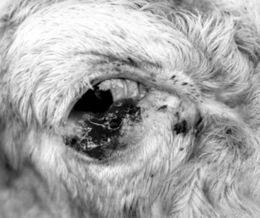

Soft tissue tumors present as progressively enlarging masses or swellings. Fibroma, fibrosarcoma, leiomyosarcoma, and leiomyoma usually occur as single masses. Neurofibroma and rhabdomyosarcoma are often multicentric. Rhabdomyosarcoma in farm animals typically arises within skeletal muscle. Cattle with neurofibromas involving large peripheral nerve trunks can develop signs of lameness and denervation atrophy.


Soft tissue tumors vary from fleshy to fatty to myxoid, depending on the tumor cell type. Cytologic evaluation will distinguish lipoma from other mesenchymal tumors. Histologic evaluation—and possibly immunohistochemistry—is needed to distinguish fibroma, fibrosarcoma, neurofibroma, leiomyosarcoma, and rhabdomyosarcoma.


Subcutaneous lipoma in farm animals is most often an isolated and discrete tumor. Other soft tissue tumors are typically locally invasive but rarely metastasize. Wide surgical excision of these tumors, when possible, could be curative.


Tumors of the Eye


Tumors involving the eyelids, conjunctiva, cornea, and orbit are relatively common in cattle and less common in other farm animal species. Ocular squamous papilloma, squamous cell carcinoma, and orbital lymphoma of cattle are most common. Ocular squamous cell carcinoma can also occur in sheep, especially those at high altitudes. Ocular dermoids are developmental anomalies present at birth that occur sporadically in calves and pigs. Ocular lymphangiosarcoma in cattle is rare.


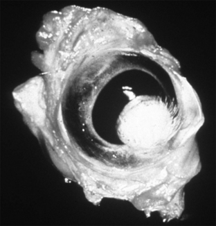
Cutaneous papillomas caused by bovine papillomavirus occur on the eyelids or corneoscleral junction of young cattle and can predispose to development of ocular squamous cell carcinoma. Bovine squamous cell carcinoma is most common in the limbal and eyelid conjunctiva but can also arise in the cornea. Squamous cell carcinoma that involves the orbit and retrobulbar tissue is much less common. Bovine ocular squamous cell carcinoma occurs in all breeds, but Herefords appear to be predisposed to this tumor. Ocular squamous cell carcinoma usually occurs in cattle over 5 years of age. Lack of eyelid pigmentation and ultraviolet light are etiologic factors, although a genetic predisposition is also suspected. Ocular squamous cell carcinoma occurs much less commonly in sheep. Lymphoma can involve the orbit of adult cattle as part of a more generalized neoplastic process associated with BLV infection. Ocular dermoid is not a true tumor but presents as an ocular mass lesion in neonatal calves and piglets. Ocular dermoid is a developmental defect in which a zone of skin, often haired, is present at birth on the cornea or conjunctiva (Figure 3-4). Ocular dermoids are most common in polled Herefords and also occur in pigs. Lymphangiosarcoma of the limbus has been reported in an 8-year-old Holstein cow. Ocular tumors have not been reported in goats.


Tumors of squamous epithelium often begin as a smooth raised plaque that progresses to form an exophytic squamous papilloma. Some ocular papillomas will spontaneously regress. Persistent papilloma can progress to noninvasive and then to invasive squamous cell carcinoma. Not all ocular squamous tumors exhibit this sequential development, and invasive carcinoma may be the first clinically noted lesion. Squamous cell carcinoma is a raised proliferative lesion that is often ulcerated and often is secondarily infected (Figure 3-5). Squamous cell carcinoma of the conjunctiva can spread to involve the globe, and corneal squamous cell carcinoma can spread to involve conjunctiva. In advanced cases, determining the initial site of malignant transformation is often impossible. Neoplastic squamous cells are seen on histologic and cytologic preparations.


Lymphoma of the orbit and retrobulbar space most often causes progressive exophthalmos of the affected eye. In dairy cattle, early signs of exophthalmos may not be noticed, as many dairy breeds normally have mild exophthalmos. When protrusion of the globe becomes severe enough to interfere with eyelid closure, rapid development of exposure keratitis occurs. The lymphoma is most often deep within the orbit, thus precluding diagnostic procedures such as biopsy or cytology. Complete physical examination for other affected organs or serologic testing for bovine leukemia virus infection are useful diagnostic procedures. Evaluation of tumor tissue by cytology or histopathology after removal of the globe reveals sheets of relatively homogeneous lymphocytes, and marked invasion and obliteration of normal architecture are seen in tissue sections.


Ocular dermoids do not enlarge, but the presence of hairs often results in irritation of the lids or cornea that can become secondarily infected. The ocular lymphangiosarcoma reported presented as a progressively enlarging subconjunctival mass at the limbus. Biopsy revealed irregular vascular channels devoid of blood, consistent with origin from lymphatics.


Ocular squamous cell carcinoma is locally invasive and destructive but metastasis is uncommon. Rarely, ocular squamous cell carcinoma in cattle undergoes spontaneous remission. Surgical removal of the affected globe and lids in cattle and sheep can be curative, although in predisposed cattle carcinoma can develop in the other eye. Other successful therapies for ocular squamous cell carcinoma include cryosurgery, hyperthermia, radiation, and immunotherapy. Removal of exophthalmic globes because of orbital or retrobulbar lymphoma should be considered only as a palliative measure to make the animal more comfortable. Complete excision of orbital lymphoma is not possible, and the involvement of other organs leads to eventual death, usually within 6 months of diagnosis. Surgical excision of dermoids is curative. Removal of the affected globe was apparently curative in the cow with ocular lymphangiosarcoma.


Tumors of the Oral Cavity and Jaw


Tumors involving oral mucosa and bones of the jaw are most common in cattle, less common in sheep, and rare in goats and pigs. Epithelial, odontogenic, vascular, osseous, and fibroblastic tumors occur.


BONE TUMORS


Tumors of bones of the jaw include odontogenic tumors, osteoma, fibroma, fibrosarcoma, and myxomatous tumors. Odontogenic tumors occur most often in cattle and sheep and rarely in pigs. Odontogenic tumors are most often evident in young to young adult animals but have also been diagnosed in older animals. Presumably, odontogenic tumors identified in adults had been present for some time before diagnosis. Tumors of odontogenic origin have a variety of histologic features that result in various—often confusing—classifications that are prone to change. Tumor types identified in farm animals include odontoma (including ameloblastic, compound, and complex odontoma), ameloblastoma, and ameloblastic fibroodontoma. Osteoma and fibroma also occur in the jaws of adult cattle. Fibrosarcoma of the jaw occurs in adult sheep grazing bracken fern. Myxomatous tumors (myxoma and myxosarcoma) occur in adult cattle.


Tumors of the jaw most often present as slowly enlarging firm to bony growths. These tumors often arise at or near tooth roots and can cause loosening and malalignment of adjacent teeth. Radiographic evaluation is useful to determine the site and extent of the tumor. Histologic evaluation is necessary to distinguish the various types of odontogenic and mesenchymal tumors that occur in the jaws of farm animals. Odontogenic tumors consist of varying elements of odontogenic epithelium with or without induction of dental or mesenchymal tissue. Biopsy diagnosis of osteoma often relies on the clinical and radiographic description of the lesion, as it is not possible to distinguish the histologic features of small samples of osteoma from those of normal bone. Myxomatous tumors are characterized by proliferation of spindle to dendritic cells in a loose myxoid stroma.


Fibroma, osteoma, and odontogenic tumors of the jaw are noninvasive and can be cured by wide surgical excision. For tumors located rostrally, mandibulectomy or maxillectomy is advised. Myxomatous tumors of the jaw, however, are typically locally invasive and difficult or impossible to completely excise. Recurrence is common, although metastasis has not been reported.

Stay updated, free articles. Join our Telegram channel

Sep 3, 2016 | Posted by in SUGERY, ORTHOPEDICS & ANESTHESIA | Comments Off on Neoplasia

Full access? Get Clinical Tree

Get Clinical Tree app for offline access