Surgery of the Sheep and Goat Digestive System

Chapter 18 Surgery of the Sheep and Goat Digestive System


Gastrointestinal surgeries in sheep and goats are not commonly performed by the private veterinary practitioner but should always be considered for individual patients of economic worth. Surgeries that are performed most commonly include drainage or resection of pharyngeal abscess (traumatic, foreign body, caseous lymphadenitis), surgeries of the forestomach (reticulorumen) and abomasum, rumen and esophageal fistula placement, correction of intestinal obstruction, or intestinal accident.


Pharyngeal trauma and subsequent abscess formation in small ruminants is a common finding. If undiagnosed, this condition can lead to cellulitis, severe tissue necrosis, dyspnea, and subsequent bloat. Radiographs, ultrasound, and endoscopic examination can help confirm and localize the lesion. If the animal is severely dyspneic and the swelling is compressing the trachea, a temporary tracheostomy is indicated. Organisms typically isolated from abscesses include: Arcanobacterium pyogenes, Yersinia pseudotuberculosis, Staphylococcus species, and Pseudomonas species. Medical therapy can be attempted, but surgical intervention may be needed if the abscess is large and the animal is symptomatic. If economically feasible, general anesthesia is warranted because of the large number of vital structures in the area, including the vagosympathetic trunk, carotid vasculature, and esophagus. Antibiotic and antiinflammatory pharmaceutical utilization should be determined by culture and sensitivity results and the severity of the lesion.


When deciding on surgical points of entry, the practitioner should always consider the location of vessels and nerves in the area. An abscess should be entered from the oral cavity whenever possible (and when attached to the oropharynx) (see Figure 10.1-5) and lanced so that it drains into the digestive system. This is best accomplished by aspirating the contents first with an 18-gauge needle and syringe. A larger gauge needle will be needed in those instances when the abscess capsule may be thick.


Either a left lateral or ventral approach is indicated for surgical drainage. The use of a blindfold in sheep and goats may accent the anesthetic effects or, in many cases, lessen the stress to the surgical patient. The surgery site is surgically prepared. It is imperative that the esophagus, carotid artery, and jugular vein be initially visualized and shielded from possible incision. Dissection should be done bluntly to avoid vital structures. Ideally the abscess is isolated and removed in its entirety. Alternatively the capsule can be sutured to the skin and the abscess marsupialized. If neither of these options is possible, the abscess can be drained by aspiration and the animal kept on long-term antibiotics.


Surgery of the Rumen


Disease of the forestomach (reticulorumen) can be fairly common in sheep and goat practice. Ruminal distention, rumen acidosis, rumen impaction, bezoar formation, and foreign body consumption (Figure 18-1) with subsequent impaction and rumenitis/reticulitis are conditions that may require surgical intervention. Advanced rumenitis generally has a poor surgical prognosis. Rumenotomy and/or trocar placement can be required to correct the other conditions.
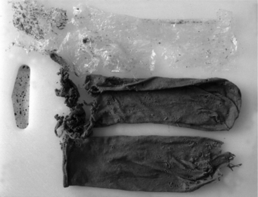


If a rumenotomy is not performed under emergency conditions, the patient should be held off feed for 12 to 24 hours before surgery. General anesthesia helps control animal movement and maintains a clean surgical field. However, if economics preclude general anesthesia use, a rumenotomy can be done with a local anesthetic and manual restraint. The practitioner should be aware that sheep and goats are highly susceptible to the toxic effects of lidocaine; therefore low volumes of diluted (1%) lidocaine should be used.


The patient is placed in lateral recumbency with the right side down and the left flank prepared for aseptic surgery. For a rumenotomy, a 15-cm vertical skin incision is made parallel and 5 cm caudal to the last rib. The underlying muscle layers can either be sharply incised or bluntly dissected along their fascia planes (“grid technique”) if a small incision is needed. Sheep and goat muscle layers are much thinner than those in cattle, and there is a more prominent cutaneous trunci muscle. Surgeons not used to small ruminants need to be careful not to make the common mistake of being too aggressive on the abdominal approach or incising over the kidneys. Once the rumen has been visualized, 10 cm should be exteriorized and sutured to the skin of the wound margin with a Lembert-type pattern around the entire incision margin. A bite is taken through the skin; then a bite is taken through the rumen (see Figure 10.3-10). This suture pattern forms a seal that prevents rumen fluid from entering the abdomen and is important to the surgery’s success. Once the suture placement is inspected and found intact, the rumen wall is incised within this margin. The surgeon should avoid traumatizing the rumen wall as much as possible.


Examination of the lumen of the rumen is now possible. Any foreign material is removed, and the cardia and reticuloomasal orifice are inspected and checked for patency. The abomasum can be palpated through the rumen wall for distention and normal location. The ventral floor of the rumen can be swept, checking for any adhesions.


It is best to close the rumen wall with two rows of continuous sutures. The second row should be an inverting pattern, such as a Cushing or Lembert, created with an absorbable suture material. Once the rumen has been closed, gloves, surgical instruments, and gowns should be changed. The wound should be flushed with copious warm saline to remove any remaining debris. Once wound cleansing has been accomplished, the rumen to skin suture should be removed and the rumen lavaged again before it is replaced into the abdomen.


Closure of the abdomen is routine, although much smaller suture material (#0 or 1) can be used than in an adult cow. Each layer is closed in a simple continuous pattern with an absorbable suture, and lavage is performed between each layer. The skin is usually closed with a nonabsorbable suture. The most ventral sutures should be placed in an interrupted fashion in case drainage of the wound is necessary. Appropriate antibiotics should be used for at least 5 days after surgery. Antiinflammatory drugs are commonly used, especially in goats (flunixin meglumine 1 to 2 mg/kg IM or butorphanol 1 mg/kg IM bid for no more than 48 hours). Clients should receive written notification of drug withdrawal times.


Rumenotomies to remove a foreign body have the most favorable prognosis. The most common items found in the rumen are plastic bags, rope, and large foreign bodies. A rumenotomy can also be performed for toxic indigestion; however, the prognosis is guarded for conditions longer than 12 hours’ duration. Medical management of these cases through intravenous fluids, electrolyte monitoring and replacement therapy, probiotics, and orogastric introduction of alfalfa meal or feed mill “fines” is usually as—or more—successful. Alkalizers* may also be helpful. Stabilization of the rumen pH and transfaunation can be important tools for successful case management. Transfaunation per os generally requires 250 to 500 ml of collection fluid 2 to 3 times daily for 3 to 5 days. This fluid should be kept anaerobic, at rumen temperature, and out of light until inoculation occurs. Ideally, the time from collection to transfaunation should be less than 30 minutes.


ABOMASAL SURGERY


Disease of the abomasum is much less common than in cattle and decidedly more difficult to manage surgically. Abomasal impaction, abomasitis, perforating abomasal ulcers, abomasal foreign bodies and abomasal emptying defect (AED) in Suffolk sheep can potentially be managed with surgical intervention. However, in most instances medical management should be attempted initially.


Generally, genetics are of extreme importance regarding abomasal impaction in sheep. Depending on an individual animal’s value and owner preference, ancillary diagnostic tools may be helpful. Ultrasound of the abomasum in the standing patient is simple and noninvasive. The abomasum is generally packed tightly with sand and ingesta. On physical examination, heart and respiratory rates are elevated. Normal rumen movements with scant feces are evident. Commonly, abomasal outflow obstruction results in hypochloremic hypokalemic metabolic alkalosis and eventually dehydration, uremia, and tachycardia. Renal excretion of NaHCO3 and KHCO3 with secondary hypovolemia occurs. Serum electrolytes should be monitored and managed before surgery. Radiology is generally of limited value and inconclusive as a diagnostic tool, although ultrasound examination may be helpful. Medical management may include the following: large volume fluid replacement, correction of electrolyte imbalance, cholinergic drugs, and IV calcium and vitamin E/selenium—all of which have been used with limited success. Abomasal impaction is much more common in goats than in sheep. Pregnancy, poor quality hay, and feeding a total pelleted diet can predispose goats to impaction. Abomasal impaction can also occur in goats confined to semidesert grazing of grassland/brush forage that contains a high percentage of awns, which form phytobezoars ranging in size from 2 to 10 cm. Patients present with inappetence, malaise, weakness, scant feces, and cranial right abdominal swelling/distension. An abomasotomy is generally corrective.


Abomasal emptying disease in Suffolk sheep commonly presents as impaction, but the etiology is different and has not been elucidated. Pregnant sheep on a diet high in concentrates are commonly affected with AED. Medical management seems to be the most common treatment course, but an abomasotomy is occasionally attempted. The flock’s genetic merit needs to be evaluated.

Stay updated, free articles. Join our Telegram channel

Sep 3, 2016 | Posted by in SUGERY, ORTHOPEDICS & ANESTHESIA | Comments Off on Surgery of the Sheep and Goat Digestive System

Full access? Get Clinical Tree

Get Clinical Tree app for offline access