Surgery of the Sheep and Goat Integumentary System

Chapter 17 Surgery of the Sheep and Goat Integumentary System


Primarily, three integumentary surgical considerations occur in sheep and goats: skinfold ablation in certain sheep breeds, tail docking or amputation, and wound care that responds to predator attacks in both sheep and goats.


Skinfold Ablation


Skinfold ablation is of primary concern in Merino and Rambouillet sheep breeds. Genetic selection for wool production over time has resulted in gross enlargement of skin folds, primarily in the neck region. Veterinary practitioners are occasionally requested to perform skin fold ablation surgery of these folds. Environmental factors such as increased temperatures, precipitation, humidity, shearing cuts, and myiasis commonly lead to moist skin fold dermatitis and secondary fly strike. Both bacterial and fungal invaders commonly populate these lesions. Mulesing (removal of skin from caudal thighs) is commonly performed in Australia and New Zealand.


As always, the owner and practitioner need to consider the value of an individual animal before under-taking this surgical procedure. In most instances, the surgical procedure is cost-prohibitive, and flock genetic selection needs to be evaluated. Procedural hemorrhage is generally minimal with rapid healing time. If possible, the surgery should be performed in young stock during the fly-free months. Skin folds are clipped and surgically prepared. Surgical incisions are made to ablate the skin fold with subsequent reapposition. Subcutaneous tissues are closed, if necessary, to appose the skin and obliterate dead space. Generally, nonabsorbable suture material is used in the skin with a simple interrupted suture pattern. Pain medication is indicated for the first 2 days. If aseptic surgical procedures are followed, antibiotic use is rarely indicated.


Predator Attack


Sheep and goats are commonly preyed upon by a multitude of carnivores. Sheep are generally attacked more often than goats, just by the nature of production practices and species temperament. Sheep and goat livestock operations near urban areas more often see attacks by domestic dogs. Dogs seem only interested in the chase of a flock or herd and are generally not hunting for food. On the other hand, wild carnivores generally kill livestock for food. It is not common to find survivors from carnivore attacks by any species other than dogs. Also, livestock carcasses are generally partially consumed and dragged to a distant site from the kill. Occasionally an owner will interrupt wild carnivores before they kill their prey, and the veterinary practitioner is called to evaluate the survivors.


Three physiologic systems seem to be of primary concern: cardiovascular (exhaustion, shock, and blood loss), penetrating wounds to the musculoskeletal system, and punctures of the gastrointestinal system. When evaluating sheep and goats maintaining heavy fleece, the initial physical examination should be thorough. Fractures and serious lacerations are extremely common in survivors. Myopathy can be a common secondary sequela. It is often difficult to thoroughly examine individuals maintaining a dense fleece.


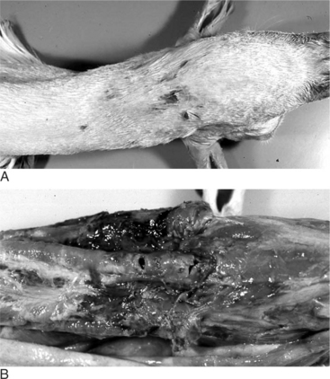
Wounds seem to focus in two areas: the ventral cervical region and head, rear limbs, and anus (Figure 17-1 A and B). Repairing fractures and lacerated ligaments in most cases exceeds an animal’s value. All but “pet” animals are many times destroyed. The veterinary practitioner should take adequate precautions to determine the most likely predatory species involved, as hostile litigation and pet destruction is a common outcome. It is common for predator (wild carnivores and domestic dogs) visits to the flock to continue. Rabies in the attacker should always be considered as a possibility, and protective gloves should be worn when dealing with saliva.


Tracheal and esophageal punctures are common and easily missed on animals maintaining a full fleece. Generally, “sucking sounds” are evident as well as subcutaneous emphysema and dyspnea. Subcutaneous emphysema may be a common sequela to all traumatic wounds from predator attack. The potential for abscesses, myopathies, tissue necrosis, nerve and ligament damage as well as secondary osteoarthritis should be discussed with owners.


Aseptic wound care is the cornerstone of patient healing. Clipping hair and wool from wounds, debriding devitalized tissues and foreign material, and cleansing the area are the initial steps of treatment. If a wound is less than 8 hours old and is easily cleaned, it may be indicated to attempt primary closure. Good aseptic technique and apposition of tissues without excessive tension gives primary closure the best chance of success. For wounds with a lot of dead space, placement of a drain may be useful.


For lacerations missing a large quantity of skin, second intention healing is indicated. Surrounding hair should be clipped, and any pockets should be drained ventrally. The wound should be cleaned often, and allowed to heal “from the inside-out.” On the same animal, some wounds may be able to be sutured while others are left open. Tetanus vaccination status should be assessed and tetanus antitoxin (500IU) is indicated in all but the most recent vaccinates. Neurologic deficits, rectal lacerations and ligament avulsion maintain a poor prognosis and generally require that the animals be destroyed.


Supportive care of the patient is extremely important for healing success. Keeping the animal warm, dry and relatively stress-free are necessary considerations. If the animal is moderately dehydrated, jugular catheter placement and subsequent fluid therapy or blood transfusion is used. If shock is extreme, corticosteroid therapy is indicated (dexamethasone 2mg/kg IV). Myopathy and orthopedic trauma require the use of nonsteroidal antiinflammatory drugs. Flunixin meglumine (12mg/kg IV or IM) or oral aspirin (100mg/kg) (use not indicated in hemorrhaging patients but fairly economical with short drug withdrawal time) therapy is also employed. Broad spectrum antibiotic therapy is indicated where slaughter is not an issue. Antibiotic choices include: penicillin (20,000IU/kg bid), florfenicol (20mg/kg IM EOD or 40mg/kg SQ) and sulfadime-thoxine (55mg/kg IV or po initially then 27.5mg/kg sid) for 5 days. Clients should always receive a written statement concerning withdrawal times and animal care in the advent of adverse drug reactions.


Consistent wound cleaning and fly control should be emphasized to the owner. Appetite and water consumption are important for the client to monitor. Animals are encouraged to stand and walk several times a day to avoid tendon contraction in the front limbs; otherwise physical therapy should be done to keep tendons stretched is indicated. Environmental stress should be minimized as much as possible.


Tail Docking


Tail docking (amputation) is usually performed in sheep (not goats) in Western nations, excluding Europe. This procedure is done to prevent fly strike, improve ram-breeding efficiency, and provide a more balanced carcass to the meat packer. Docking is typically performed in the first two weeks of the lamb’s life. However, it is not uncommon for the veterinary practitioner to dock the tail on a mature ewe overlooked at a younger age. The 1- to 2-week-old lamb is docked with little or no hemorrhage. Techniques include the following: hot chisel, electric hot docker, emasculator, Burdizzo, elastrator, and blade amputation. The tail is amputated at the mid to distal limit of the caudal tail fold found on the ventral aspect of the tail. The remaining “dock” or stump should cover the anus. The recent producer trend of extremely short tail docks has led to an outbreak of a number of secondary health problems, most notably rectal prolapse.


An assistant holding the patient upside down with each hand locking the respective front and rear limbs usually accomplishes restraint of a lamb. Surgical clipping and cleaning is not generally employed if the tail is clean and free of fecal debris. Tetanus antitoxin (250IU SQ) is indicated if previous dam vaccination has been ignored. The practitioner should not recommend elastrator/rubber bands. They function through ischemic necrosis, and tetanus is a common sequela.


Amputation of a mature tail is a serious surgical procedure. General anesthesia is recommended, but many practitioners use physical restraint accompanied by either a caudal epidural perfusion block or local infusion with 2% lidocaine. Postsurgical analgesia is always indicated. The tail is surgically clipped and cleaned. A tourniquet is applied at the base of the tail for hemostasis. The mature tail is amputated at the mid to distal limit of the caudal tail fold found on the ventral aspect of the tail. The loose skin should be retracted proximally before incision and amputation to allow adequate reapposition. The skin is incised in a “V” pattern to permit easy closure. The vertebra are either incised interdigitally or crushed and cut with an emasculator for added hemostasis. Closure is completed with nonabsorbable 0 to 2-0 suture material in a simple interrupted or horizontal mattress suture pattern. Tetanus prophylaxis (antitoxin 500IU SQ), seasonal fly control, and appropriate antibiotics are given after surgery.


17.1 Caprine Dehorning


When deciding whether to dehorn a goat, one must consider several factors. Goats with horns pose a threat to other animals and the people that work with them. Some goat breeds cannot be registered or shown until they are dehorned. Goats without horns are less destructive to farm facilities and are less likely to become entangled in fences. In addition, dehorning can be combined with descenting in males. However, dehorning is not appropriate for all goats. Goats that range or are kept on tethers should be allowed to keep their horns as a defense mechanism. In addition, dehorned bucks may be less able to compete with horned herdmates for breeding purposes and dehorning in adulthood may have secondary complications that include delayed healing, decreased milk or sperm production, and possibly death.


Given the complications and costs associated with dehorning, select breeding for polled goats would seem to be advantageous. However, goats have a dominant polled gene closely linked to an infertility recessive gene. Goats homozygous for the polled condition are less fertile because of conditions such as sterile intersex females and a predisposition toward sperm granulomas in males. Breeding programs should account for this possibility, and polled goats should not be interbred to avoid these complications.


Disbudding


Removal of horn buds in young goats is most appropriately termed disbudding and should be performed within the first week of life. European breeds of buck kids should be disbudded between 3 and 5 days of age while doe kids should be disbudded between 5 and 7 days of age. The horns of Nubian kids grow more slowly than European breed kids, which allows disbudding in Nubians to be delayed until two weeks of age. Some goat kids are polled and will not need to be disbudded. Polled goats can be recognized by a single whorl of hair on top of their heads, whereas horned goats have a whorl of hair over each horn bud.


RESTRAINT AND ANESTHESIA


Several methods of restraint and anesthesia are available for disbudding kids. Although some prefer to use physi-cal restraint alone, others use a combination of physical restraint, local anesthesia, sedation, and general anesthesia. If physical restraint is used alone, a goat disbudding box can be useful. The boxes are roughly 24 inches long, 18 inches tall, and 7 inches wide, with a lid and small opening at the front of the box for the kid’s head. Kids dehorned with physical restraint alone resume normal behavior immediately after the disbudding process has been completed, which some think is justification enough for this method.


The cornual branch of the infratrochlear and lacrimal nerves innervate the horn bud of the goat. A subcutaneous line block along the dorsomedial rim of the orbit blocks the cornual branch of the infratrochlear nerve. The site to block the cornual branch of the lacrimal nerve is located halfway between the lateral canthus of the eye and the posterior edge of the horn along the cornual ridge behind the supraorbital process. Local anesthesia is performed in kids by injecting 1 milliliter of a solution (1 milliliter of 2% lidocaine diluted with 3 milliliters of sterile water) into each of four sites required to block the two horn buds (Figure 17.1-1).
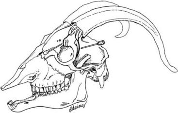


In the field, xylazine (0.06mg/kg IV) can be used to provide sedation. A 2% to 3% halothane gas can be used to anesthetize kids when dehorning is performed in an office. The high oxygen content of the halothane gas may make the goat’s hair prone to combustion in the presence of a hot iron; therefore it must be removed before the iron is applied.


SURGICAL TECHNIQUE


Heat cautery is the most commonly used technique to disbud kids. A 200-watt dehorning iron with a ¾ to 1-inch tip is used for heat cautery. Lower wattage dehorning irons do not get as hot and must be applied to the head longer to burn the same degree as the higher wattage models. The hair over the horn bud should be clipped before disbudding to improve visualization and decrease the amount of smoke inhaled by the person performing the disbudding.


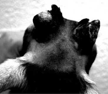
Once the dehorner has become cherry red, it should be applied to the horn bud for 3 to 4 seconds while being rocked around the bud. The head should be allowed to cool before reapplying the iron for another 3 to 4 seconds. Two applications of the iron should be adequate to completely destroy the horn corium, and this is assured if a ring of copper-colored skin that encircles the horn bud and cannot be scraped off with a fingernail has formed. The circle of skin inside the ring should be burned as well. Buck kids require a larger ring of burnt skin than doe kids do, and bucks can also be descented at this time by burning an additional crescent of skin caudomedial to each horn bud (Figure 17.1-2).


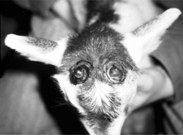
The most common mistakes associated with using heat cautery include inadequate burning that leads to scur formation (Figure 17.1-3) and excessive burning that leads to heat meningitis. The frontal bone’s thinness and the absence of a frontal sinus at the age kids are disbudded make them prone to heat meningitis. Signs of heat meningitis include unresponsiveness and an inability to nurse. Treatment with antibiotics, antiinflammatory agents, supplemental heat, and tube feeding may allow some affected kids to recover from this condition.


Stay updated, free articles. Join our Telegram channel

Sep 3, 2016 | Posted by in SUGERY, ORTHOPEDICS & ANESTHESIA | Comments Off on Surgery of the Sheep and Goat Integumentary System

Full access? Get Clinical Tree

Get Clinical Tree app for offline access