Chapter 20 Necropsy
Description And Interpretation of Gross Observations
Lesions encountered on gross examination can be described in terms of the following characteristics1:
Weights of individual organs may aid diagnosis. For example, liver weight may vary depending on the presence or absence of toxic hepatopathy, and thyroid weight may be increased in cases of goiter. Volume of gastrointestinal content or fluid in compartments also may provide reliable objective data. Many common veterinary diagnoses, such as trauma, lightning strike, and certain types of bloat, can be made only by gross examination (Figure 20-1, A and B). In such instances, submission of tissues for histopathologic examination and culture to a diagnostic laboratory, without appropriate history and descriptive gross observations, may result in no diagnosis.
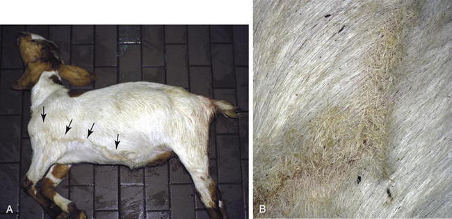
Table 20-1 lists commonly encountered systemic pathologic processes along with their characteristic necropsy lesions and possible causes.2–8 Table 20-2 lists gastrointestinal diseases that may require gross examination for diagnosis.6,8–13 Table 20-3 lists “pseudolesions” that may be confused with pathologic changes4 (Figure 20-2). Astute clinical observation and field necropsy constitute the first line of defense against exotic or reportable livestock diseases in the United States. Table 20-4 lists necropsy lesions that may be observed in goats and sheep infected with reportable agents.14–32
TABLE 20-1 Selected Systemic Pathologic Processes: Necropsy Findings and Potential Causes
| Pathologic Process | Possible Lesions | Cause(s) |
|---|---|---|
| Cardiogenic shock | Pulmonary congestion and edema, jugular vein distention, large liver with “nutmeg” pattern, generalized edema, anasarca, ascites, submandibular edema, hydropericardium, hydrothorax, hydroperitoneum, heart lesions (cardiomegaly, myocarditis, hydatid cysts, myocardial abscess, hemorrhage/pallor, white streaks in myocardium, morphologic defect, myocardial mineralization)2 | Hypoxia, ventricular tachycardia, and other arrhythmias, cardiomyopathy, congenital defect, hypertension, endocarditis (chronic infections, long vascular blood catheter use), bacterial or viral infection, nutritional deficiency (selenium, copper), cardiotoxic substance or plant (e.g., vitamin D, rhododendron/azalea), monensin toxicity, electrocution or lightning strike, obstruction of blood flow, fluid overload3 |
| Obstruction of blood flow | Pericardial fluid or exudate accumulation, thrombus in vena cava or lung | Thrombosis, congenital (aortic stenosis), cardiac tamponade (traumatic reticulopericarditis, ruptured coronary artery), neoplasia, bloat (abdominal or rumen distention), liver failure |
| Hypovolemic shock | Pale or swollen kidney, contracted spleen | Hemorrhage, fluid loss (dehydration) |
| Hemorrhage | Extravasation of blood, pallor of tissues (lungs, mucous membranes), watery/thin blood, observation of blood in body cavity, contracted spleen | Enteric hemorrhage, predator attack, ruptured viscera (spleen, uterus, abomasa), ruptured liver from dystocia, ruptured uterine or ovarian artery, abomasal ulceration, rupture of aorta, ruptured aneurysm4 |
| Fluid loss (dehydration) | Sunken eyes, reduced skin elasticity | Diarrhea, burn injury, acute intestinal obstruction, electrolyte imbalance, acid-base imbalance5 |
| Maldistributive shock | Reduced circulating blood, volume pooling in capillaries, congested spleen, pulmonary edema | Endotoxemia, septicemia (gram-positive or -negative bacteria septicemia), rapid reduction of body fluid (removal of ascitic fluid),10 neurologic dysfunction, anaphylactic shock, neurogenic shock, septic shock2,5 |
| Anaphylactic shock | ||
| Neurologic shock | Nonspecific gross lesions that are similar to cardiogenic shock | Trauma, electrocution, lightening strike, fear and stress2 |
| Septic shock | Petechiae, injected sclera, ecchymoses, large swollen lymph nodes, splenomegaly, fibrinopurulent exudates in pericardial space or joint spaces or adhered to meninges5 | Bacteremia |
| Endotoxemia (toxic or septic shock) | Lesions may be same as for toxemia5 | Lipopolysaccharide cell wall of gram-negative bacteria |
| Toxemia (toxic shock) | Pale or enhanced liver parenchyma, enlarged kidneys, enlarged adrenal glands, hemorrhage of myocardium, coagulopathy, increased vascular permeability, congestion, distended intestines, presence of glucose in urine6 | Bacterial toxins (enterotoxemia), tetanus, inorganic toxin, plant toxins, nitrate toxicity, snake bite, xenobiotic toxicity (e.g., from tilmicosin),4 urea toxicity |
| Edema | Fluid accumulation in tissues, pleural cavity, or abdominal cavity5 or in subcutis or ventrum | Congestive heart failure, obstructed venous return, endotoxemia, anaphylactic shock, vasculitis, obstruction of lymph flow5 |
| Hypoxia | Nasal discharge, pulmonary changes, cyanosis | Anemia, cardiac failure, pneumonia, rhinitis, pulmonary edema, infectious disease, toxicity (cyanide anemia, nitrate or carbon monoxide toxicity), metabolic disease (pregnancy toxemia), neoplasia, pneumothorax, tracheal collapse, allergic pneumonitis, hyperthermia, trauma (diaphragmatic hernia, tight collar)4 |
| Hypothermia | Pulmonary congestion, generalized congestion | Cold stress, exposure to rain or wind; may occur after shearing4 |
| Hyperthermia | Congested tissues, petechial hemorrhages in mucous membranes, subcutis; hyperemia of skin | Infection, high environmental temperature |
| Liver failure | Altered liver size (small or large), altered liver texture (firm or friable), ecchymoses, enhanced lobular pattern | Hepatitis, hepatopathy |
| Renal dysfunction | Urine obstruction, swollen pale kidneys, dilated ureter or calyces | Urolithaisis, glomerulonephritis, nephritis, nephropathy, |
| Coagulopathy | Petechial or ecchymotic hemorrhage, subcutaneous hemorrhage, pooling of blood in spaces or lumina of organs | Disseminated intravascular coagulation, reduction in vitamin K–dependent clotting (toxic plant ingestion, rodenticide toxicity), snake envenomation, fungal toxins (Aspergillus, Fusarium), liver flukes (Fasciola hepatica), bacterial infection7 |
| Emaciation | Decreased subcutaneous, pericardial adipose tissue, muscle atrophy, poor hair or wool quality, oral disease, changes in liver parenchyma | Dental disease, protein-energy malnutrition,5 micronutrient deficiency, gastric foreign body, chronic enteritis, chronic liver disease, infectious (prion disease, retroviral infection, chronic septicemia, chronic parasitism), plant toxicities, neoplasia, amyloidosis8 |
TABLE 20-2 Digestive Tract Diseases: Necropsy Findings and Potential Causes (see Chapter 5)
| Digestive Disease | Possible Lesions | Cause(s) |
|---|---|---|
| Esophageal choke | Distended esophagus with thinned or hemorrhagic wall | Inadequate mastication, large/firm food item, foreign body, injury from stomach tube, injury to tissue surrounding esophagus |
| Rumen acidosis | Distended rumen with porridge-like content and “fermented” odor, rumen pH <5.5,9,10 rumen epithelium hyperemic and sloughs readily,11 dehydration (sunken eyes), chronic (scarred rumen lining, liver abscesses)11; urine pH 5.0-6.0; packed cell volume >35%6,9 | Rapid rumen fermentation with ingestion of highly digestible carbohydrates (corn, oats, wheat, barley, breads, fruits, beets, potatoes, others)9 |
| Rumen distention, impaction | Distended rumen, esophageal “bloat line,” edema in hindquarters, dry scant feces | Protein-energy or micronutrient malnutrition,10 ingestion of foreign body (plastic bags),8 dehydration, legume, ingestion of high-fiber/low-digestibility diet, sand ingestion, consumption of horse feed9 |
| Frothy bloat | Esophageal “bloat line,” flattened liver, diaphragm rupture; edema in hindquarters, hindquarters blanched11 | Legume consumption (alfalfa), lush cereal-grain pastures, high-grain diets |
| Free gas bloat | Rumen distended with gas, esophageal “bloat line” | Inhibition of eructation (rumen malfunction, esophageal obstruction, newly introduced grain diet, neurologic function impaired, hypocalcemia, endotoxemia, pain, peritonitis, some pharmaceuticals (e.g., xylazine)9,12 |
| Traumatic reticuloperitonitis | Peritonitis, pericarditis, draining tracts from chest cavity | Ingestion of wires, needles |
| Abomasal impaction | Abomasum enlarged with rumen-type contents that are dry and doughy | Poor-quality roughage, foreign body obstruction (phytobezoar, trichophytobezoar), protein-energy malnutrition, abomasal atony, emptying defect in Suffolk and Dorset sheep, dysautonomia9,11 |
| Abomasal bloat in juveniles10 | Distended abomasa with milk content | Associated with large quantities of milk replacer diet,10 consumption of hay mixed with feces13 |
| Abomasal rupture6 | Feed content in peritoneal space, tear in greater curvature of abomasa accompanied by hemorrhage in abomasal wall | Abomasal impaction, abomasal bloat, abomasal atony |
| Abomasal torsion6 | Hemorrhage and congestion at the site of the torsion | Gas accumulation and displacement10 |
| Intestinal volvulus | Hemorrhage, gas distention, mesenteric rent (tear) | Gas accumulation in intestine |
| Intussusception | Intestinal telescoping most commonly observed at ileocecal valve | Associated with intestinal mass in adults, enteritis in young, Oesophagostomum in sheep9 |
| Intestinal ileus | Dilated intestine with watery content | Multiple-system disease |
| Intestinal atresia, | Failure of intestinal segment to form, first week of life | Congenital defect |
| Cecal volvulus/torsion of root of mesentery | Distention of forestomachs, abomasa, and intestine | Gas accumulation and displacement9,10 |
| Rectal prolapse | Protrusion of rectal tissues | Diarrhea in lambs and kids; dietary imbalance, urolithiasis, grazing lush pasture; secondary to chronic coughing, short tail docking, growth implants, rabies, atresia ani9 |
| Atresia ani | The rectum is not patent | Congenital defect |
TABLE 20-3 Pseudolesions Observed at Necropsy
| Pseudolesion | Necropsy Findings |
| Bird damage | Loss of eyes, damage to anus |
| Euthanasia barbiturate | Soft, discolored dark tissue at injection site |
| Gunshot euthanasia (owner may not inform vet concerning method of euthanasia) | Hemorrhage and fractures, bullets |
| Liver mortis-postmortem | Settling of blood in dependent parts of body4 |
| Digestive overflow-postmortem | Rumen contents in bronchi, nasal cavity4 |
| Rumen distention after death (“postmortem bloat”) | Rectal or vaginal prolapse |
| Terminal lesions associated with death process | Tracheal froth from cardiac hemorrhages |
| Congenital melanosis | Dark pigmentation of meninges, other tissue4 (see Figure 20-2) |
| Normal placenta features mistaken for lesions | Necrotic membrane tip, hippomanes, amniotic plaques (see Figures 20-17 and 20-18) |
| Autolysis | |
| Diaphragm rupture (postmortem) | Free gas in rumen combined with autolysis; mimics rupture from bloat or trauma |
GI, Gastrointestinal.
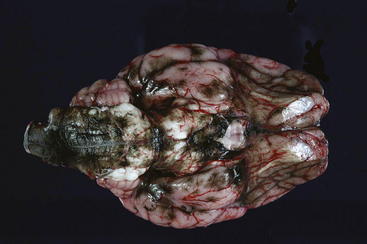
TABLE 20-4 Reportable Diseases of Worldwide Significance or Foreign to the United States: Endemic Distribution and Necropsy Lesions
| Disease* with Causative Pathogen When Known | Endemic Distribution | Necropsy Findings |
| PRION DISEASE | ||
| North America, Europe, Asia, Australia14 | Emaciation, loss of wool or hair, traumatic skin lesions caused by rubbing14 | |
| VIRAL DISEASES | ||
| Africa, Asia, Europe, South America, previously in North America15 | High morbidity, low mortality; usually affects younger animals; lesions in goats and sheep are mild or nonapparent Small vesicles or erosions on the dental pad, lips, gums, or tongue; vesicle or erosion on the coronary band or in the interdigital space; decreased milk in mammary gland; gray or yellow streaking in the myocardium; abortion is possible16 | |
| South America, Central America, North America15 | Less frequent in sheep and goats, high morbidity, rarely causes death; vesicles on tongue, gums, lips, coronary bands, and possibly teats; secondary bacterial mastitis16 | |
| Worldwide except some Pacific islands, Australia17 | No gross lesions | |
| Africa, Middle East, Indian subcontinent15 | Emaciation; inflammatory lesions in GI tract from oral cavity to colon; dehydration, fecal staining; stomatitis involving inside of lower lip and adjacent gum, cheeks near commissures and free portion of tongue; lesions possible on hard palate, pharynx, upper third of esophagus, rumen, reticulum, omasum, abomasa; Peyer’s patch necrosis, congestion at ileocecal valve and cecocolic junction, “zebra stripe” congestion in colon Respiratory lesions: erosion and petechiae of nasal mucosa turbinates, larynx, and trachea; bronchopneumonia | |
| Africa, Middle East, Asia, previously Europe and South America15 | Digestive tract lesions: small gray foci on oral mucosa that coalesce, gray necrotic epithelium that sloughs and leaves red erosion; erosions on gums, lips, hard and soft palate, cheeks, and base of tongue; erosion of esophagus/omasum, congestion and edema of abomasum; Peyer’s patch necrosis, fibrinous exudates on intestinal mucosa, edema and/or luminal blood in cecum and colon; congestion of colonic ridges—“tiger” or zebra striping, swollen or edematous lymph nodes16 | |
| Africa, Middle East, Asia17 | 1- to 3-cm-diameter macules on skin, particularly in groin, axilla, perineum; papules on mucosal membranes of nose, mouth, mammary glands, vulva, or prepuce; nasal discharge from rhinitis; mastitis, multiple necrotic foci of mucous membranes, enlarged lymph nodes; papules on mucosal surface of abomasum, rumen, large intestine, tongue, hard and soft palate, trachea, and esophagus; pale areas on surface of kidney, liver, and testicle; hemorrhagic enteritis, multifocal firm areas in lungs, abortion possible16,17 | |
| Scotland, England, Ireland17 | No characteristic gross lesions17 | |
| Africa, Middle East15,16 | Hepatic necrosis; enlarged friable, soft, red to yellow-brown liver; petechial to ecchymotic hemorrhage; patchy congestion, small gray-white 1- to 2-mm foci in liver parenchyma, petechiae and ecchymoses in mucosa of abomasa, dark chocolate-brown abomasal contents; edema and hemorrhage in wall of gallbladder and hepatic lymph nodes16 | |
| Africa16 | Hemorrhagic and catarrhal gastroenteritis, soiling of hindquarters with mixture of blood and feces; hemorrhages or congestion in longitudinal folds of the abomasum distal ileum, ileocecal valve, cecum, and colon; congestion or hemorrhage in the cecum and colon appearing as longitudinal striations of the mucosa (“zebra striping”); colon contents may be watery and blood-tinged, with hemorrhages in serosa of colon, submucosa of gallbladder, epicardium, and endocardium; nasal discharge, prominent lymph nodes, enlarged spleen; abortion is possible with numerous hemorrhages in tissues, edema, and hemorrhage of fetal membranes16 | |
| Africa, Asia, Australia15 | Encephalitis with no gross lesions, stillborn, weak or aborted fetus, atrophy of skeletal muscle, tendon contraction, arthrogryposis, hydranencephaly, torticollis, scoliosis, brachygnathism15 | |
| Africa18 | Liver in fetus and newborn is enlarged and orange-brown, with multifocal pinpoint white areas; icterus often present; hydranencephaly, microcephaly, arthrogryposis, hydrops amnion in ewes16,18 | |
| Worldwide in mostly tropical and subtropical climates, Europe, North America15 | Vascular injury, consumption coagulopathy, generalized edema, hyperemia, hemorrhages, erosions, ulceration of upper gastrointestinal tract (oral cavity, esophagus, forestomachs), subintimal hemorrhages in pulmonary artery, pulmonary edema, pleural effusion, pericardial effusion, edema in facsial planes, necrosis of papillary muscle in left ventricle; embryonic or fetal death, fetus: cavitating encephalopathy, hydranencephaly16 | |
| Europe | No characteristic gross lesions; possible leptomeningeal hyperemia, brain edema16 | |
| Worldwide | Emaciation, chronic polysynovitis, degenerative joint disease, hyperplasia of synovium, enlarged lymph node, diffusely firm lungs (interstitial pneumonia), mastitis17 | |
| Africa, North America, South America, Europe, Asia17 | ||
| Africa, North America, South America, Europe, Asia17 | Solid raised foci in lungs, pulmonary neoplasia in anterior-ventral regions and diaphragmatic lobes, excessive froth in bronchi, enlarged bronchial and mediastinal lymph nodes with occasional metastasis, secondary pneumonia, pulmonary abscess, pleuritis17 | |
| North America, South America, Europe17 | No characteristic gross lesions, in purities cases damage to skin, subcutaneous edema, pulmonary congestion and edema, hydropericardium, endocardial hemorrhages17 | |
| BACTERIAL DISEASES | ||
| Worldwide | Absence of rigor mortis, rapid gaseous decomposition; dark tarry nonclotting blood at orifices, widespread ecchymoses, blood-stained fluid in body cavities, hemorrhagic enteritis, splenomegaly with “blackberry jam” consistency19 | |
| Asia, Australia, Middle east, Africa, Caribbean, South America (tropical regions)20 | Thick exudates from eyes and nose, multiple abscesses in subcutis associated with lymph nodes and abscesses in many organs (lungs, spleen, liver), abscess may contain green-tinged pus, aortic lesions, joint effusions, cloudy meninges20 | |
| Africa | Hydropericardium, pericardial fluid that is reddish to straw-colored, decreased packed cell volume, orange-yellow serum, hydrothorax, pulmonary edema, ascites, edema of lymph nodes, mediastinal edema, froth in the trachea, subendocardial petechial hemorrhages, subtle swelling of the brain, partial brain herniation, possible splenomegaly16 | |
< div class='tao-gold-member'> Only gold members can continue reading. Log In or Register to continue
Stay updated, free articles. Join our Telegram channel
Full access? Get Clinical Tree
 Get Clinical Tree app for offline access

| ||