48 Multiple canine mast cell tumours
CASE HISTORY
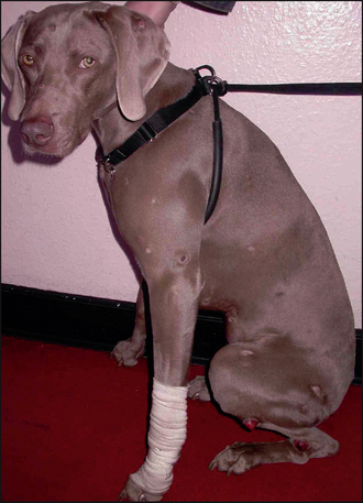
This dog presented with the following history:
CLINICAL EXAMINATION
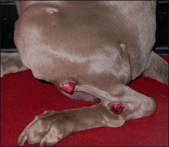
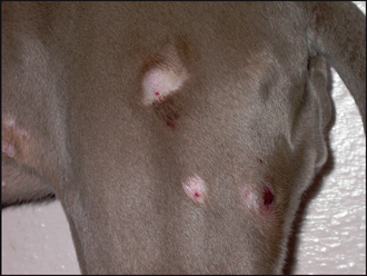
In this case the findings were:
CASE WORK-UP
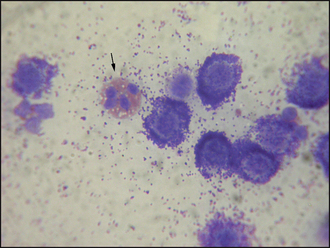
The first step with any nodular disease is to perform fine-needle aspiration. Except in the case of poorly differentiated tumours, the diagnosis of MCTs is usually easily made on cytological examination of fine-needle aspirates. Diff Quik® or Rapi-Diff® are appropriate stains for cytological examination. Mast cells appear as small to medium-sized round cells, the nuclei are centrally placed within the cell, giving a ‘fried egg’ appearance, and the cytoplasm usually contains abundant dark staining granules (see Chapter 2). Eosinophils are frequently encountered. NB: Mast cell granules may be difficult to visualize in poorly differentiated MCTs.
The advantages of fine-needle aspiration over excisional biopsy are that it is inexpensive, it can be performed with the animal conscious and, in the case of mast cell tumour, is usually diagnostic. Additionally, an effective first surgery is most likely to result in a cure and prior fine-needle aspirates allow for effective surgical planning. The major limitation of fine-needle aspiration is that it is not possible to predict the behaviour of the tumour; histology is much more useful in this respect.

Figure 48.4 Mast cells with both intracellular and extracellular granules. Also shows an eosinophil (arrow).
Bearing in mind these limitations, the following tests were carried out:
Stay updated, free articles. Join our Telegram channel

Full access? Get Clinical Tree


