Fig. 47.1
Light micrographs of HE- and PAS -stained liver tissues by various preparation methods. With HE-stained CB liver tissues at surface area, congested erythrocytes are observed (a: small arrows, b: large arrows), but open sinusoidal space and flowing erythrocyte s are observed in slightly deeper areas (a: arrowheads, c: small arrows, CV central vein ). Eosin-stained congested erythrocytes (d: large arrows) and weak hematoxylin-stained large hepatocyte nuclei are observed by the QF liver tissues (d: small arrows). HE-stained hepatocyte cytoplasm along cell membranes is observed by the IF liver tissue (e: arrows). PAS-reaction product s are strongly and homogeneously observed throughout the hepatocyte cytoplasm in the surface area of CB specimen (f: small arrows and arrowheads, h: small arrows, h: large arrows; open sinusoid ). In slightly deeper area of the CB specimen, hepatocellular cord with strong PAS-reaction products is clearly observed (g: arrows). Strong but heterogeneous PAS-reaction products are observed in the QF hepatocyte cytoplasm (i: arrows). Weakly and unevenly PAS-reaction products are observed in the IF hepatocytes (j: small arrows; weak, large arrows; strong). (a, f) and (g) Bars: 50 μm. (b, c, d, e, h, i) and (j) Bars: 20 μm (Adapted from Fujii et al. [18])
47.3 PAS -Staining of Mouse Livers by Different “Biopsy” Techniques
For preparing the common “biopsy” and immersion-fixed specimens for light microscopy , extracellular and intracellular components were easily leaked out or moved with mechanical compression forces or dehydration and embedding steps [19]. In the CB specimens, PAS -reaction product s were strongly and homogeneously recognized in the cytoplasm of hepatocytes (Fig. 47.1f–h). However, they were heterogeneously observed in the conventional quick-freezing (QF) liver hepatocyte cytoplasm (Fig. 47.1i, small arrows). On the other hand, by conventional immersion fixation and dehydration samples, PAS-reaction products were weakly observed in most of hepatocyte cytoplasm (Fig. 47.1j, small arrows). So, the CB technique would be a useful tool for examining soluble component s .
47.4 Morphology of Living Mouse Livers by the CB Technique for SEM
By in vivo cryotechnique , the sinusoidal erythrocytes could be cryoimmobilized in vivo with various shapes in blood vessel s , as reported before [10, 11]. At the initial contact surface of freeze-fractured CB liver tissues, some erythrocytes were compactly localized in the hepatic sinusoids and the Disse’s space was not clearly open (Fig. 47.2a). To the contrary, open hepatic sinusoids and flowing erythrocyte s were clearly observed in slightly deeper areas with Disse’s spaces (Fig. 47.2b). The morphology of freeze-fractured hepatocytes showed nuclei, many globular mitochondria, and endoplasmic reticulum in their cytoplasm (Fig. 47.2c). At higher magnification, freeze-fractured mitochondria (M) and endoplasmic reticulum (ER) were clearly observed in the hepatocytes (Fig. 47.2c, inset). The Disse’s spaces were covered with sinusoidal endothelium and hepatocyte surface with microvilli (Fig. 47.2c, asterisk). In some areas of Disse’s spaces, hepatocyte cell membranes were slightly separated from each other, which reached deeper near bile canaliculi (Fig. 47.2c, small arrows). The CB samples applied to freeze-fracture technique for SEM, functioning three-dimensional structure, would be examined in living animal livers.
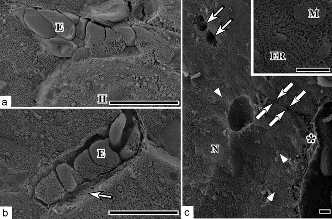
Fig. 47.2


Scanning electron micrographs of the CB specimens. At the surface areas, aggregated erythrocytes , which show no biconcave discoid shape s , are observed, but Disse’s spaces are hardly recognized (a: E erythrocyte , H hepatocytes ). In slightly deeper areas, erythrocytes are separated and Disse’s spaces are clearly observed (b: E erythrocyte, Arrow Disse’s space). Some globular cell organelles and Disse’s spaces are observed under the normal blood circulation (c: arrowheads; globular cell organelles, asterisk and large arrows; Disse’s spaces, N nucleus). The Disse’s space reaches near bile canaliculi (c: small arrows). Inset: At higher magnification, abundant endoplasmic reticulum (ER) and freeze-fractured mitochondria (M) are clearly observed. (a) and (b) Scale bar = 10 μm. (c) Scale bar = 1 μm (Adapted from Fujii et al. [18])
Stay updated, free articles. Join our Telegram channel
Full access? Get Clinical Tree
