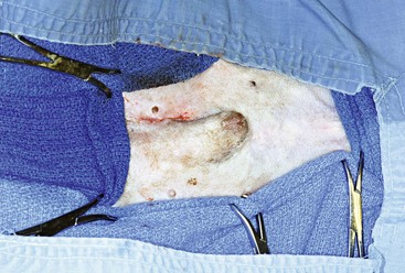
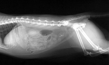
Chapter 199 Urocystolithiasis occurs commonly in dogs and cats. Although medical dissolution protocols are available for some types of uroliths (e.g., struvite, urate, and cystine), alternative methods of managing urocystoliths often are pursued because of failure of dissolution, inability to attempt dissolution, or lack of dissolution protocols (e.g., for calcium oxalate stones) (Bartges and Lane, 2003). Open cystotomy is the conventional means for removing urocystoliths; however, minimally invasive techniques such as transurethral cystoscopy and laser lithotripsy in female dogs and cats (Adams et al, 2008; Lulich et al, 2009) and laparoscopically or cystoscopically assisted cystotomy in male and female pets (Libermann et al, 2011; Rawlings et al, 2003; Runge et al, 2011) are becoming more commonplace. Minilaparotomy-assisted cystoscopic retrieval maximizes the advantages of both and avoids the equipment and expertise required for laparoscopy. Rather than use a laparoscope to assist in urinary bladder identification and entry, which requires additional specialized equipment and establishment of pneumoperitoneum, minilaparotomy-assisted cystoscopy relies on the surgeon to isolate the apex of the urinary bladder and temporarily fasten it to the linea alba, which allows cystoscopy and stone removal to be performed through a small stab incision in the urinary bladder wall. Indications for minimally invasive urocystolith retrieval are similar to those for open surgical cystotomy and include (1) medically nondissolvable urocystoliths (e.g., calcium oxalate or urate uroliths associated with portovascular anomalies), (2) client or patient factors that preclude medical dissolution, and (3) a need to remove the urocystoliths more rapidly than is possible with medical dissolution. Additionally, these minimally invasive procedures may be performed multiple times for a patient with recurrent urocystoliths; adhesions to the urinary bladder serosal surface appear to occur less commonly than with open cystotomy. Minilaparotomy-assisted cystoscopy typically is performed in male dogs and cats but can be performed in female dogs and cats when transurethral cystoscopy is not feasible. Minilaparotomy-assisted cystoscopy is most useful for patients with large stones that would require extended time to fragment with laser lithotripsy, for patients with large numbers of stones that would also make lithotripsy or open cystotomy time consuming, and for patients whose size precludes lithotripsy (see Web Chapter 70). In addition to allowing urolith retrieval, minilaparotomy-assisted cystoscopy also is useful for obtaining biopsy specimens of urinary bladder mucosa, evaluating whether hematuria is renal in origin by observing urine flow from ureteral orifices, and facilitating ureteral stent placement in certain circumstances. With the transvesicular placement of a rigid cystoscope, the proximal urethra, including the prostatic urethra in male dogs, may be examined in an antegrade fashion. Most of the remaining urethra can be visualized using an appropriately sized flexible endoscope. Before the procedure, a thorough history should be taken and a complete physical examination should be performed. Collection of a minimum data set including results of complete blood cell count, biochemical analysis, and urinalysis is indicated. Additional testing may include an aerobic bacteriologic culture of urine collected by cystocentesis, liver function testing if urate urocystoliths are suspected, and testing of adrenal function or calcium metabolism if calcium oxalate uroliths are likely. Abdominal survey radiography should be performed (Figure 199-1), but ultrasonographic examination or contrast studies also may be required to determine the locations of all uroliths and their impact on the urinary tract. The patient is placed under general anesthesia after appropriate premedication and is positioned in dorsal recumbency. If necessary, a urinary catheter is inserted in the urethra and urethroliths are retropulsed into the urinary bladder. Before final preparation of the abdominal wall, this urinary catheter is removed. The ventral abdominal wall is clipped, prepared, and draped as for standard laparotomy. The preputial sheath is flushed with 0.05% chlorhexidine solution for two minutes (Neihaus et al, 2011). A sterile urethral catheter then is inserted in male dogs and cats. The size of the catheter depends on the size of the patient: a 3.5 Fr catheter in a male cat, a 5 Fr to 10 Fr catheter in a male dog. Female dogs and cats usually do not require urinary catheterization to facilitate isolation of the bladder. The urinary bladder is distended with sterile fluid such as lactated Ringer’s solution or normal saline so that the urinary bladder is palpable through the abdominal wall. An incision is made through the skin on the ventral midline over the urinary bladder. In male dogs the incision is made just cranial to the preputial reflection or, if the urinary bladder is displaced caudally, it may be made in the right or left peripreputial area (Figure 199-2). The skin incision is 2 to 3 cm in length. The subcutaneous tissue is incised and the linea alba is identified. A stab incision is made through the linea alba and lengthened using Mayo scissors. Figure 199-2 Final appearance of minilaparotomy-assisted cystotomy skin incision made in the right peripreputial area in a 6-year-old castrated male bichon frise.
Minilaparotomy-Assisted Cystoscopy for Urocystoliths
Indications
Preprocedure
Procedure
![]()
Stay updated, free articles. Join our Telegram channel

Full access? Get Clinical Tree


