Management of Rectal Tears

CHAPTER 101 Management of Rectal Tears


The rectum extends from the pelvic inlet to the anus and is a storage chamber for feces. It is approximately 30 cm long and is divided into a peritoneal and a nonperitoneal section. The peritoneal rectum is a continuation of the small colon, and this determines its position, which is most commonly in the left dorsal aspect of the pelvic cavity.


The small colon and peritoneal portion of the rectum are structurally similar, and the rectal vasculature arises from the internal pudendal artery and vein via a continuation of the small colon mesentery, the mesorectum. The nonperitoneal portion of the rectum varies in length because the distance between the peritoneal reflection and the anus is inconsistent. The nonperitoneal portion of the rectum is longer in old horses and fat horses, and the distance from the anus does not differentiate between the peritoneal and nonperitoneal sections. The nonperitoneal section of the rectum lacks a serosal covering and is attached to surrounding tissues by connective tissue and muscular bands. It is attached to the pelvic diaphragm, and ventrally it is attached to the vagina in mares and to the urethra in stallions. The most caudal portion transitions into a flask-shaped dilation called the ampulla recti. The mural structure varies from the longitudinal muscle fiber bundles in the small colon and peritoneal rectum into loosely associated longitudinal muscle bundles at the ampulla recti.


Feces are evacuated by passing from the ampulla recti through the 5-cm-long anus. The circular muscle of the rectum ends at the anus as the internal anal sphincter, which is under involuntary control, and the external anal sphincter consists of striated muscle that is under voluntary control.


DIAGNOSIS


Clinical signs seen in horses with a rectal tear vary with the severity of the tear and duration of the problem. If a tear occurs during rectal palpation, the veterinarian may feel a sudden release in pressure, cessation of straining, an improved ability to palpate the abdominal structures, and possibly the presence of blood on the rectal sleeve. A horse with a rectal tear may strain to defecate, pass hemorrhagic feces, and become colicky. Depression, endotoxemia, and peritonitis may follow within a few hours.


Establishing the severity of a tear is of vital importance because it dictates treatment and prognosis. This is best achieved by preventing further straining using intravenous (IV) xylazine hydrochloride (0.1 to 0.2 mg/kg) in combination with IV butorphanol tartrate (0.1 mg/kg). Lidocaine (50 mL of 2% lidocaine per rectum) can be used to relax the rectum; however, IV butylscopolamine bromide (0.3 mg/kg) is more effective at reducing rectal pressure. Once the horse is sedated, an epidural anesthetic can be administered (0.2 mg/kg xylazine hydrochloride and 0.22 mg/kg mepivacaine chloride diluted in saline to 10 mL) to enable exploration of the tear.


The rectum should be evacuated of all feces. Digital inspection with a lubricant-covered, ungloved hand is the most effective means of determining the extent and severity of a rectal tear. Typically the redundant rectal mucosal folds prevent direct view of the injury, although some tears may be inspected using a vaginal speculum or an endoscope. Endoscopic evaluation of the rectum is commonly performed at referral facilities to facilitate therapeutic decision-making.


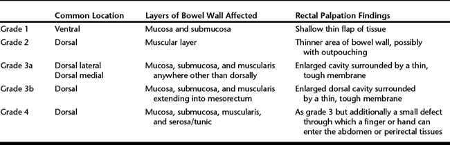
Tears are graded according to the involvement of the layers of the bowel wall as initially reported by Arnold and colleagues in 1978 (Table 101-1). Tears can be located at any distance from the anus; however, most are 25 to 30 cm from the anus at the junction of the rectum and small colon. This area is considered to be predisposed to injury because the blood vessels penetrate the intestinal wall acutely and directly rather than gradually, therefore causing weakening in the dorsal aspect of the wall. Iatrogenic tears are most commonly longitudinal, but idiopathic tears may be transverse.


After confirmation of a rectal tear, it is advisable to perform abdominocentesis for cytologic analysis and culture, although changes in the peritoneal fluid might not be evident immediately.


TREATMENT


Once the injury has been confirmed, the owner should be promptly informed of the treatment options and prognosis. Immediate care should focus on preventing progression of the tear, facilitating healing, and treating the secondary consequences such as peritonitis, endotoxemia, shock, and laminitis.


Grade 3 and 4 Tears


Grade 3 and 4 tears are acute life-threatening emergencies, and transportation to a referral institution is recommended. Grade 4 tears associated with gross fecal contamination of the abdomen frequently justify euthanasia. However, appropriate emergency care should be initiated before transportation if there are any doubts regarding the depth of the tear. The importance of appropriate immediate treatment of a grade 3 or 4 tear cannot be overemphasized because of its influence on prognosis.


Once the rectum has been evaluated and measures have been taken to counteract straining, antimicrobial drugs should be initiated immediately. Antimicrobial therapy should be broad-spectrum and should include drugs with activity against anaerobes (potassium penicillin, 22,000 units/kg IV every 6 hours; gentamicin sulfate, 6.6 mg/kg IV every 24 hours; and metronidazole, 15 to 25 mg/kg PO every 8 hours). NSAIDs (flunixin meglumine, 1.1 mg/kg IV every 12 hours) and tetanus prophylaxis should also be administered. Although the importance of minimizing contamination within the tear is undisputed, there is some controversy regarding the most appropriate method of achieving this. Packing the rectum with an antiseptic-covered tampon has been advocated. The tampon should be positioned in the rectum so that its cranial end is 10 cm cranial to the tear. A tampon can be constructed by tying a length of stockinette at both ends and filling it with cotton soaked in diluted Betadine solution. Lubricating the tampon with a medical lubricant before insertion may avoid mucosal adherence and facilitate extraction. The aboral end of the tampon is secured to a length of umbilical tape, which can be brought out through the rectum for subsequent removal. The rectum may be closed with towel forceps or a purse-string suture to prevent expulsion of the tampon.


Rectal packing may predispose to worsening of the tear through manual trauma and an increase in intrarectal pressure, especially if the horse strains. If a rectal tampon is not used, it may be sufficient to ensure the rectum is fully evacuated and that the horse is treated with parasympatholytics such as butylscopolamine bromide (0.3 mg/kg IV) and epidural anesthesia, which in combination should decrease straining and facilitate fecal passage.

Stay updated, free articles. Join our Telegram channel

May 28, 2016 | Posted by in EQUINE MEDICINE | Comments Off on Management of Rectal Tears

Full access? Get Clinical Tree

Get Clinical Tree app for offline access