CHAPTER 162 Management of Bladder Uroliths
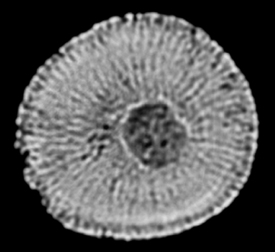
The most common type of uroliths in the horse are those composed of calcium carbonate, but a few are composed of calcium phosphate. The former are rough and friable, and the latter are smooth and solid stones. A urolith forms over a nidus of cellular debris and will readily form over a foreign body such as a fragment of nonabsorbable suture. Management of a male horse with a bladder urolith is one of the most challenging surgical problems involving the equine urinary system. Over their career, most equine clinicians will diagnosis only a few of these calculi, and the difficulties of treatment and need to manage and monitor affected horses on a long-term basis after surgery make a lasting impression on both owner and veterinarian. Combined with the technical challenges of urolith removal is the dearth of evidence-based information on postoperative clinical management that will best minimize recurrence. Identification of the specific components of primary inhibitors and promoters (e.g., Tamm Horsfall glycoprotein, nephrocalcin, pyrophosphate, and uromucoid) of calculus formation in the horse awaits research. Elucidating the mechanisms of failure of the natural inhibitors of calculus formation may hold the key to improved approaches for diagnosis and prevention.
DIAGNOSIS
Transrectal palpation is the initial step in confirming the presence of a bladder urolith. Transrectal examination of the bladder is facilitated by the use of sedation, epidural anesthesia, or administration of N-butylscopolammonium bromide (0.2 to 0.3 mg/kg, IV). Clinical signs of urolithiasis are seen with uroliths 1 to 10 cm in diameter; although a solitary urolith is typical, multiple calculi can develop. Drainage of the bladder improves detection of smaller uroliths. Endoscopic examination should be performed before surgical intervention is undertaken to examine the bladder mucosa and urolith surface. Urine should be collected for urinalysis and culture, and each ureteral opening should be evaluated for normal appearance, location, and function. Because they are not a typical feature of bladder urolithiasis, biopsy should be taken of any full-thickness erosive lesions of the bladder mucosa. When a bladder urolith is detected, ultrasound examination of the urinary tract should be recommended to detect kidney uroliths and to assess renal architecture if there is biochemical evidence of renal dysfunction. Bladder urolithiasis should be clearly differentiated from sabulous cystitis (see Current Therapy in Equine Medicine, ed 5, page 833).
PREOPERATIVE PREPARATION
Details of preoperative preparations vary with surgical approach, but the following is a discussion of measures that should be observed regardless of the approach. The horse should be withheld from full feed to reduce intestinal tract volume. This improves the surgeon’s ability to operate in or around the bladder by reducing intestinal contact and pressure on and near the bladder. The duration of feed withholding or intake reduction (by feeding low bulk-high caloric feedstuffs) will depend on the volume of ingesta in the digestive tract at the outset and rate of passage. Mild exercise promotes intestinal evacuation. The goal is to achieve a concave shape to the paralumbar fossae, which generally requires 24 to 72 hours. Appropriate antimicrobials should be administered if bacteria are identified by cytology or culture. If no bacteria are detected, administration of antimicrobials and anti-inflammatories should be reserved for the perioperative period. Most horses have negative urine culture results, although it is not uncommon to obtain microbial growth from the nidus of a urolith (Figure 162-1).


