Management of Atrial Fibrillation

CHAPTER 41 Management of Atrial Fibrillation


Atrial fibrillation (AF) is the most important cardiac rhythm disorder affecting performance in horses. The prevalence is about 0.5%, and there is no known sex or age predilection. AF is an atrial tachyarrhythmia that is frequently found in the absence of other cardiac pathology, especially in large breeds. In ponies, AF is encountered only as a result of severe cardiac disease.


MECHANISMS AND PATHOPHYSIOLOGY


Atrial fibrillation is the result of a reentry mechanism in the atrial myocardium that occurs as a result of tissue heterogeneity in refractoriness and conduction. Different models have been described to explain the reentry phenomenon. The well-known multiple wavelet model assumes that during AF multiple electrical wavefronts move chaotically through the atrial myocardium. For AF to be self-sustaining, a critical minimum number of wavefronts must coexist in the atria. More recent information from human medicine has revealed AF to originate from rapidly firing foci or from small reentry sites (spiral waves or rotors), usually in the left atrium or the ostium of the pulmonary veins. These sites act as a continuous source of electrical waves that spread through the atrial myocardium.


When both a trigger to start the arrhythmia and a substrate to maintain it are present, AF develops. The trigger can be one or more atrial premature beats that initiate reentry. Myocardial damage, myocardial stretching, electrolyte disorders, or systemic disease can cause these premature beats. The substrate, the atrial myocardium, must be sufficiently large, have a short refractory period, have differences in the length of refractory period, have slow conduction velocity, or have structural obstacles or lesions or any combination of these conditions. Predisposing factors for AF include high vagal tone, high sympathetic tone, large atria such as those in large breeds or horses with atrial dilatation, atrial stretch as occurs during exercise, valvular disease, and myocardial lesions.


Occasionally, AF is short-lived and terminates spontaneously, usually within the first 24 to 48 hours, in which case it is called paroxysmal AF. This form of AF can be a cause of reduced performance in slow-finishing Thoroughbreds. It is thought that strenuous exercise triggers initiation of AF by stretching the atrial myocardium, leading to atrial premature beats and electrophysiologic changes that are exacerbated by electrolyte disturbances, whereas the myocardium does not support sustained AF after exercise because of its size, structure, and electrophysiologic properties.


In most horses, however, AF does not terminate spontaneously once initiated because the size and properties of atrial myocardium are a suitable substrate for maintaining the arrhythmia. In addition, immediately after AF develops, electrophysiologic and contractile remodeling ensue, leading to further stabilization of the arrhythmia. AF becomes permanently established and will not terminate without treatment.


During AF, continuous and chaotic self-sustaining electrical activity persists in the atria at a rate of approximately 300 to 500 pulses per minute. Because of vagal tone, the atrioventricular (AV) node blocks most of these electrical pulses, and the final ventricular rate remains normal when the horse is at rest. Within days to weeks after initiation of AF, atrial contractile function is almost completely lost, a process that is reversible after restoration of sinus rhythm. Although atrial contraction contributes as much as 15% to 20% to ventricular filling, loss of atrial contractility is not related to clinical signs at rest because passive filling is sufficient to maintain cardiac output. However, during exercise, when heart rate increases, the atrial contribution to filling becomes more important. In addition, exercise results in a predominance of sympathetic tone whereby the AV node conducts too many pulses to the ventricles and results in a disproportionate tachycardia during exercise. Both factors can result in reduced exercise tolerance, which, in the absence of other cardiac disease, is mainly observed during vigorous exercise.


CLINICAL PRESENTATION


As is true in other species, AF in horses may develop secondary to an underlying cardiac problem, such as mitral valve regurgitation or congestive heart failure. The primary cardiac disease makes the atria more susceptible to AF because of atrial dilation, myocardial stretch, and fibrosis. In the event of these changes, antiarrhythmic treatment will be more likely to fail, and even if AF can be converted to sinus rhythm, recurrence is likely because of the predisposing heart disease.


Most frequently and in contrast to other species, however, horses (but not ponies) develop AF without underlying cardiac disease or with only minor underlying disease. This condition is called lone or primary AF. Treatment of horses with this form of AF is rewarding because of the good success rate for cardioversion and lower rate of recurrence.


Clinical signs depend on whether the horse has concurrent cardiac or noncardiac disease. Initiation of AF in horses with progressively developing cardiac failure generally results in aggravation of clinical signs and an increase in heart rate (usually to around 60 beats/minute).


Horses that have AF without underlying cardiac disease usually have a history of reduced performance at high-level exercise. Epistaxis may develop during exercise. Occasionally a brief period of ataxia or distress may be observed during fast work. In noncompeting horses, AF may be an incidental finding.


On auscultation of the heart, AF is characterized by an irregularly irregular rhythm and loud first heart sound. Careful auscultation easily distinguishes between AF and second-degree AV block because during AF the rhythm is more irregular, the first heart sound is louder, an unexpected early beat will always be heard, and an atrial sound is absent, especially during a long pause. After slight excitation, the arrhythmia remains. The heart should be auscultated carefully immediately after exercise because the irregularity in R-R intervals during tachycardia is less pronounced as a result of shortened diastolic time. In addition, postexercise sinus arrhythmia commonly develops in normal horses and can easily be confused with AF. Horses with secondary AF may have clinical signs related to the predisposing disease. However, a horse with lone AF should not have signs of heart disease at rest. Arterial pulse quality is usually variable. Inspection of the jugular veins may reveal intermittent filling of the veins during a long diastolic pause. This should be distinguished from pathologic venous pulsation that can be seen with right-sided heart failure.


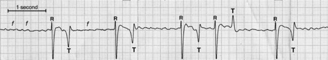
Diagnosis of AF should be confirmed by means of an electrocardiogram (ECG), which will be characterized by normal QRS morphology and irregular R-R intervals, coarse undulations of the isoelectric line (f waves), and absence of P waves (Figure 41-1). Because of aberrant conduction, a shortly coupled QRS complex might have a different T-wave morphology, which should not be mistaken for a ventricular premature beat. The presence of ventricular premature beats suggests an electrolyte disorder or diffuse myocardial disease and warrants further investigation.


May 28, 2016 | Posted by in EQUINE MEDICINE | Comments Off on Management of Atrial Fibrillation

Full access? Get Clinical Tree

Get Clinical Tree app for offline access