45 LMN paresis and paralysis
Brachial plexus avulsion
CLINICAL EXAMINATION
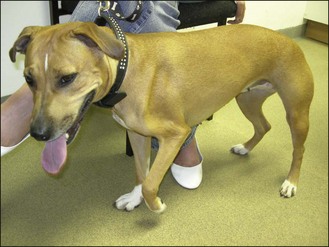
The dog was alert and ambulatory with a non-weight-bearing left forelimb. Severe generalized atrophy of the left fore was present (Fig. 45.1). The left elbow was held flexed. Muscle contracture prevented extension of the right carpus, elbow and shoulder. The left cutaneous trunci reflex was absent when either side was stimulated. The left pupil failed to dilate fully in darkness. Pain perception was absent in the radial and ulnar nerve distributions but was present in the distribution of the musculocutaneous nerve.
Stay updated, free articles. Join our Telegram channel

Full access? Get Clinical Tree


