55 Torticollis INTRODUCTION Scoliosis describes a lateral curvature of the spine. It may be congenital due to a wedge-shaped malformation of vertebral bodies (hemivertebrae). Acquired scoliosis occurs secondary to syringomyelia or hydromyelia from unequal tone in or sensory input from the spinal musculature. Kyphosis The dorsal deviation of the thoracic vertebral column is seen as an arching of the back. It commonly accompanies back and abdominal pain. Vertebral malalignment from hemivertebrae or vertebral fracture or luxation are less frequent causes. Lordosis is a downward curvature of the lumbar spine with exaggeration of the normal dip between the scapulae and the hip (dipped back, swayback). It can be due to hemivertebrae or vertebral fracture/luxation. Torticollis also known as ‘wry neck’, results from an involuntary unilateral contraction of the neck muscles causing torsion (twisting) of the neck. Head rotation (head tilt) with laterocollis (turn to one side), retrocollis (dorsiflexion), but usually not antecollis (ventroflexion) is seen. It may be secondary to an acquired cervical spine or spinal cord lesion, or muscle spasm from local or higher CNS origin. SIGNALMENT A 9-year-old male Maltese terrier. CASE PRESENTING SIGNS A twisted neck and inability to walk. CASE HISTORY The dog had tried to jump onto a chair to steal a hamburger but was restrained by a short lead and fell backwards, hitting the ground. The dog had remained immobile in a twisted position since the accident 2 hours prior to referral. CLINICAL EXAMINATION 
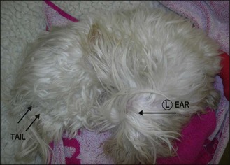
The dog was obtunded and in lateral recumbency with an almost 90° turn of his head and neck to the right. The left eye was visible and had a menace response, a normal direct PLR in a medium sized pupil. The left fundus was normal. There was no strabismus or spontaneous nystagmus in the left eye. The right eye was hidden by the abnormal posture (Fig. 55.1). Figure 55.1 Torticollis. Left ear (arrow). Tail (double arrows). Only gold members can continue reading. Log In or Register to continue You may also needUMN: Vertebral malformationUMN: Spinal fractureLeaks, dribbles urineAtaxia: Spinal ataxiaUrinary incontinence – an introductionRetroperitoneal diseaseUMN: Spinal empyemaHypocalcaemia Share this:Click to share on Twitter (Opens in new window)Click to share on Facebook (Opens in new window)Click to share on Google+ (Opens in new window) Related Tags: Saunders Solutions in Veterinary Practice Small Animal Neurology Sep 3, 2016 | Posted by admin in SMALL ANIMAL | Comments Off on Torticollis Full access? Get Clinical Tree