Thomas J. Divers
Leptospirosis
Leptospirosis is caused by a highly invasive spiral bacterium of the genus Leptospira. The infectious agent is capable of infecting both humans and animals. Less is known about leptospirosis in horses than in any of the common domestic animals except cats. On the basis of DNA-DNA reassociation studies, genus Leptospira is classified into 13 named species and 4 genomospecies, several of which contain both pathogenic and nonpathogenic serovars. Serovars, which are based on the older phenotype classification, are sometimes classified as causing host-adapted infection or incidental host infection. Host-adapted strains seldom cause clinical disease in their maintenance host, infection and shedding are prolonged, and the serologic response following infection is relatively low. Conversely, incidental host serovars are more likely to cause clinical disease in a nonmaintenance host, be associated with a marked serologic response following infection, and be shed only briefly by the host.
In North American horses, Leptospira interrogans serovar Pomona type kennewicki is the prominent incidental (pathologic) serovar, and foxes, opossums, raccoons, deer, and skunks are believed to be the most common maintenance hosts for this serovar. In Europe, important equine strains are Leptospira kirschneri serovar Grippotyphosa, strains duster (Western Europe) and moskva (Eastern Europe). In South America, L interrogans icterohaemorrhagiae and copenhageni are important strains. L interrogans serovar Bratislava is considered by most researchers to be the host-adapted serovar of the horse. This belief is met with some controversy, however, because horses may have high serum titers of antibodies against serovar Bratislava, and some investigators believe it to be pathogenic in horses.
Clinical Syndromes
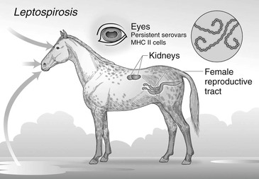
Pathogenic Leptospira infections in the horse appear to have organ trophism for the kidney, eye, or female reproductive tract (Figure 42-1). Infection may result in placentitis and abortion, neonatal jaundice, acute renal failure or hematuria, and most important, uveitis. More recently, thrombocytopenia, acute pulmonary hemorrhage with respiratory distress, fever, and acute renal failure were reported in five 1- to 3-month-old foals.
Reproductive Tract Infection
Leptospira interrogans serovar Pomona abortions may account for approximately 13% of bacterial abortions in mares in endemic regions, although incidence varies considerably between years. The reason for the yearly variation in incidence of abortions is not clear. Serovar Pomona type kennewicki is responsible for most of the Leptospira abortions in North America, but serovars Grippotyphosa and Hardjo have also been reported. Most abortions occur after 9 months of gestation, and, rarely, a live foal may be born ill from leptospirosis. Moreover, infected fetuses carry Leptospira in the placenta, umbilical cord, kidney, and liver. Lesions include placentitis that does not involve the cervical star. Macroscopic lesions are edema and areas of necrosis in the chorion. Microscopic lesions include necrosis and calcification of the placenta. Placental disease may result in the mare developing hydrallantois. Macroscopically, the fetal liver may have yellow discoloration. Liver disease is caused by multifocal necrosis and giant cell hepatopathy. Tubulonephrosis and interstitial nephritis may be detected in the kidney of the aborted fetus. Inflammation of the umbilical cord, funisitis, may be recognized by diffuse yellowish discoloration. It is unknown whether abortion results because of the placentitis, funisitis, or fetal infection or the effects of all three. Although more than one mare on a farm may abort because of Leptospira infection, abortions in epidemic areas are unusual. Aborting mares and other recently infected horses are believed to shed L interrogans serovar Pomona in the urine for approximately 2 to 3 months. A small number of horses on a farm with one or more Leptospira abortions may develop uveitis weeks later.
Acute Renal Failure
Occasionally, Leptospira pomona causes fever and acute renal failure in horses. The kidneys become swollen as a result of tubulointerstitial nephritis, and urinalysis may reveal hematuria and pyuria without visible bacteria. On rare occasions, multiple weanling or yearling horses may be affected with fever and acute renal failure following L pomona infection.
Recurrent Uveitis
The most important clinical disease associated with L interrogans serovar Pomona infection in adult horses in North America and L kirschneri serovar Grippotyphosa in Europe is recurrent uveitis (see Chapter 150). Two distinct ocular diseases appear to be associated with L interrogans serovar Pomona infection: the most common is equine recurrent uveitis (ERU), but immune-mediated keratitis may also occur. The strong association between ERU and L interrogans serovar Pomona dates back to the early 1950s with a general belief that it was an immune-mediated disease involving antibody against certain Leptospira antigens, specifically the LruC outer membrane protein, which cross-reacts with tissues of the lens, cornea, and possibly retina.
Since 2000, numerous scientific publications have confirmed detection of live Leptospira organisms in the uveal tissue, aqueous, or vitreous fluid of horses with recurrent uveitis. High concentration of antibody against L interrogans serovar Pomona in the aqueous humor, compared with serum titers, also suggests persistent local antigenic stimulation. Survival of the organism in the face of high ocular antibody indicates an absence of cells or molecules (e.g., complement) involved in bacterial clearance, suggesting an ocular immune privilege similar to that of the central nervous system. Recurrent episodes of the disease may be related to a Th17 response of autoreactivity following mimicry and intermolecular or intramolecular epitope spreading, or both.
Genetic factors are likely involved in the disease process, helping to explain why only some horses infected with Leptospira develop uveitis. Appaloosas are thought to be genetically predisposed. Recurrent uveitis is the most common cause of blindness in horses. The prevalence of ERU is unknown, but reports suggest that 1% to 7.6% of horses will develop the disease during their lifetime. It is probable that some cases of ERU are not associated with Leptospira infection, and this may vary by geographic region. In some regions, more than 50% of ERU cases are associated with persistent ocular infections with Leptospira. Leptospira-associated uveitis may cause corneal, anterior chamber, and posterior chamber disease. Therefore clinical findings may vary from corneal edema, clinically quiet retinal lesions observed on funduscopic examination, and most dramatically recurrent and progressive painful uveitis. The chronic disease of the globe may cause cataracts, retinal degeneration, or even glaucoma.
Stay updated, free articles. Join our Telegram channel

Full access? Get Clinical Tree


