Michelle C. Coleman
Urolithiasis
1Dormia Stone Dislodger, V. Mueller Co., McGow Park, IL.
Urolithiasis is an uncommon disease in horses, with a reported prevalence of 0.11% of equine admissions to veterinary teaching hospitals and 7.8% of diagnoses of urinary tract disease. Urolithiasis is more common in males than in females, likely attributable to the longer and less distensible male urethra, which impedes passage of calculi. A breed predisposition has not been recognized. Uroliths are most commonly identified in the urinary bladder (60%), although they may also develop in the kidneys (12%), ureters (4%), and urethra (24%). In approximately 9% of affected horses, uroliths have been detected in more than one location.
The etiology of calculi formation is not completely understood. Urolith formation begins with mineralization around a nidus under conditions favoring crystal growth. In horses, urine stasis and supersaturation of urine, large quantities of calcium, and increased uric acid concentration support nidus formation. The nidus can consist of necrotic debris, mucoproteins, leukocytes, desquamated epithelial cells, or a foreign body such as suture material. Once a urolith forms, enlargement occurs by aggregation of calcium carbonate crystals in the form of spherules or precipitation of crystals on the surface of the stone.
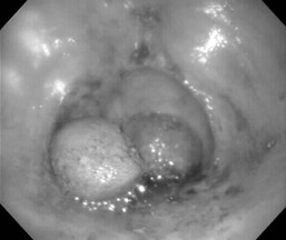
Two types of urolithiasis are recognized. Type 1 is the most common in the horse and involves uroliths that are yellow-green in color, are spiculated on the surface, and consist of a variety of hydrated calcium carbonate salts. Type 2 uroliths are less frequently observed and are white-gray in appearance with a smooth surface. The composition of type 2 uroliths is mostly calcium carbonate salts, but they may also contain magnesium and phosphorus. Type 2 uroliths have a harder composition, making them more difficult to break down and remove than type 1 uroliths.
Clinical signs, diagnostic approaches, and treatment options can vary significantly depending on the site of urolith formation and degree of obstruction.
Nephrolithiasis and Ureterolithiasis
Renal and ureteral calculi are less common than cystic calculi. Horses with cystic calculi may also have calculi in the upper urinary tract. Nephroliths typically form around a nidus as a result of underlying renal disease such as pyelonephritis, renal tubular or papillary necrosis, or neoplasia. It has been speculated that damage to the renal medulla caused by prolonged use of nonsteroidal antiinflammatory drugs may result in an increased risk for nephrolith formation.
Horses with nephrolithiasis or ureterolithiasis may remain asymptomatic, with upper urinary tract stones only identified incidentally at necropsy. Clinical signs become apparent if obstructive disease or renal disease develops. Nonspecific clinical signs of obstructive disease include colic, stranguria, and hematuria; signs consistent with uremia include poor performance, decreased appetite, lethargy, and weight loss.
Diagnosis of renal or ureteral calculi is usually made on the basis of rectal palpation or ultrasonographic examination. Rectal findings may include an enlarged kidney or dilated ureter; calculi can occasionally be palpated in an enlarged ureter. Large calculi may be sonographically identified within the kidney or ureter. Stones smaller than 1 cm in diameter may be difficult to locate. Ultrasonographic findings that support upper urinary tract obstructive lithiasis include dilation of the renal pelvis or proximal ureter, or hydronephrosis. A quantitative urine culture should be performed in all horses with nephrolithiasis or ureterolithiasis to assess possible concurrent urinary tract infection.
Surgical removal of the calculi is most effective in horses that do not have chronic renal disease. Surgical options include nephrectomy, nephrotomy, and ureterolithectomy using ventral celiotomy and paralumbar approaches. In the absence of azotemia, nephrectomy is preferred to eliminate any associated upper urinary tract infection or risk for recurrence. Less invasive surgical options include removal with a basket stone dislodger1 inserted through a vestibuloureteral approach, electrohydraulic lithotripsy, and extracorporeal shock-wave lithotripsy. A recent report described successful removal of ureteral calculi with uterine biopsy forceps used in a standing flank approach. This method is low cost, requires minimal specialized equipment, and allows easy access to the ureters.
Cystic Calculi
Cystoliths are the most common type of urolith in horses. A common observation with cystolithiasis is hematuria associated with exercise. Other common clinical signs include dysuria, stranguria, oliguria, and pollakiuria. Tenesmus, tail swishing, stamping of the hind feet, and dribbling of urine have been reported. Diagnosis of cystoliths is made on the basis of history, rectal examination, ultrasonographic (Figure 108-1) and endoscopic examinations, and urinalysis. Cystic calculi can often be detected during rectal palpation. Stones are most easily palpated when the bladder is empty, and palpation may be facilitated with the use of sedation, epidural anesthesia, or administration of N-butylscopolammonium bromide (0.2 to 0.3 mg/kg, IV). Single uroliths are most common, but multiple calculi may be present. Endoscopic examination should be performed before surgical removal for evaluation of the bladder mucosa and the urolith’s appearance. Both ureters should be identified and evaluated for appearance, location, and function. Urine should be collected for urinalysis and quantitative culture. The entire urinary tract should be assessed for the presence of other uroliths.
< div class='tao-gold-member'>
Stay updated, free articles. Join our Telegram channel

Full access? Get Clinical Tree


