Edmund K. Hainisch, Sabine Brandt
Equine Sarcoid
With a prevalence ranging from 1% to 12%, sarcoids represent the most common tumor type in equids, including horses, donkeys, zebras, and mules. Sarcoids are benign yet locally aggressive skin tumors that can arise anywhere on the animal’s body, but there is some predilection for the head, neck, and ventral body surfaces. Sarcoids may manifest as single or multiple lesions. Classified according to gross appearance, six clinical types can be distinguished: mild-type occult and verrucous lesions, and more aggressive nodular, fibroblastic, mixed, and malevolent sarcoids.
Occult sarcoids usually manifest as flat, hairless, roughly circular lesions that may contain small cutaneous nodules or be mildly hyperkeratotic (Figure 99-1, A). They frequently develop around the eyes and mouth, on the neck and chest, or on other relatively hairless body sites. Occult sarcoids usually grow slowly or remain quiescent for several years. Alternatively, they may progress to become verrucous lesions (Figure 99-1, B) or convert to aggressive fibroblastic lesions (Figure 99-1, D). This is especially the case when lesions become subject to injury, a phenomenon that is likely a result of the viral etiology of disease. Verrucous sarcoids are often encountered in the ears, axilla, groin, and sheath areas. They are of rough, wartlike appearance, are poorly circumscribed, and can affect extensive skin areas. These lesions usually grow slowly, but they can rapidly progress to fibroblastic lesions when traumatized. The predilection sites of nodular sarcoids include the ocular region, chest, groin, and sheath. Lesions appear as firm, pedunculated or sessile, subcutaneous nodules that are initially covered by normal-appearing skin. On spontaneous or trauma-mediated tumor growth, the overlying skin becomes thinner, adherent to the nodule, and eventually ulcerated, and conversion to the fibroblastic type of sarcoid takes place.
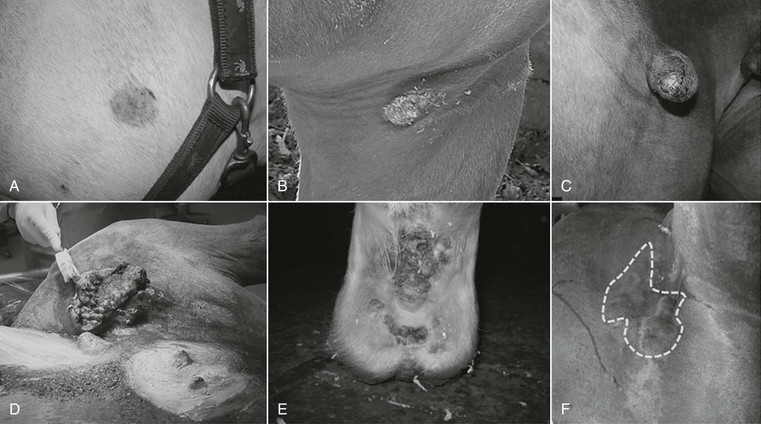
A fibroblastic sarcoid is characterized by a fleshy, ulcerated appearance and possible serum exudation (see Figure 99-1, D). Fibroblastic sarcoids commonly affect the groin and the eyelids, may develop spontaneously as primary lesions, or may result from accidental or iatrogenic injury of other sarcoid forms at any anatomic site. Malevolent sarcoids (Figure 99-1, F) are rare, and constitute the most aggressive form of the disease. This form of sarcoid frequently develops on the head, along the neck, or on the elbow or medial thigh area. Lesions usually involve a fibroblastic lesion from which extensions radiate under the skin. In contrast to all other types of sarcoids, malevolent lesions firmly attach to the underlying fascia and muscle tissue. The skin itself may appear normal over a wide portion of a malevolent sarcoid; the sarcoid infiltrates local lymphatic vessels and thus usually encompasses multiple cords of tumor masses.
Mixed sarcoids present as tumor conglomerates, which can comprise any of the previously mentioned types in any combination. They frequently occur on the face and eyelid, groin, elbow, and thigh, and usually emanate from long-standing lesions or result from ineffective tumor treatment.
The type and number of lesions affecting any given horse can vary. Some horses may bear a single quiescent lesion for many years, and others can develop several hundred sarcoids of multiple types. At present, it is accepted that injury coinduces sarcoid development and promotes progression from mild-type to more severe types and from single to multiple lesions. Accordingly, early diagnosis and effective treatment are crucial. Histologically, sarcoids are characterized by fibroblastic proliferation with epidermal hyperplasia and hyperkeratosis. Dermal fibroblasts frequently form a “picket fence” configuration by perpendicular arrangement along the dermoepidermal junction, and they generally present in various unusual configurations, including characteristic whorls and other tangled patterns.
Although geldings and young horses, as well as certain breeds such as Quarter Horses, Appaloosas, and Haflingers, seem to be at higher risk for developing sarcoids, all equids have the potential to develop sarcoids. In addition, data from different horse breeds indicate that certain equine leukocyte antigen haplotypes may genetically predispose to the condition.
Causal Association of Bovine Papillomavirus Infection with Sarcoid Development
Papillomaviruses are small icosahedral viruses that consist of a capsid harboring a circular, double-stranded DNA genome. They are usually highly host specific and require the environment of epithelial cells to complete a productive life cycle. In other cell types, notably dermal fibroblasts, they reside as circular DNA episomes, which replicate and continue to express regulatory and transforming proteins. The latter are responsible for the hyperproliferation and immortalization of infected cells, the conversion from normal to neoplastic cells. In cattle, infection by bovine papillomaviruses of types 1, 2, or both (BPV-1, BPV-2) commonly induces benign warts, which spontaneously regress. In a few cases, and notably upon ingestion of carcinogenic substances contained in bracken fern, lesions may progress to neoplasia. In addition to the disease induced in cattle, BPV-1 and BPV-2 are the major causative agents of equine sarcoid. Infection of equids by these bovine viral types constitutes one of the rare examples of papillomaviruses infecting two different species. The earliest evidence of a causal association between BPV-1/2 and equine sarcoids was obtained by inoculation experiments in the early twentieth century. BPV-1 prevails in Europe and, BPV-2 prevails in the western United States. Bovine papillomavirus 1 and 2 genomes and transcripts, as well as corresponding proteins, are detectable in almost 100% of lesions, and various in vitro and ex vivo studies have revealed some aspects of the virus-induced pathogenesis of disease. Importantly, BPV infection is not restricted to the lesions but also involves the entire integument and peripheral mononuclear blood cell subsets of sarcoid-affected animals. This has crucial implications on sarcoid diagnosis and therapy. The presence of BPV-1 or BPV-2 infection in clinically unremarkable horses remains an open issue.


