39 Lens – introduction
The lens can be involved in primary disease (e.g. cataract or lens luxation) or can exhibit pathological change secondary to other ocular disease such as uveitis and glaucoma. An understanding of its anatomy and physiology is necessary to appreciate its role in ocular disease.
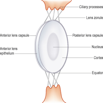
The lens is a biconvex, transparent and refractive structure suspended by zonular fibres, situated behind the iris, dividing the anterior and posterior segments of the eye. The lens consists of a capsule, lens epithelium and the lens fibres. The lens is subdivided into a central nucleus and surrounding cortex, and these can be further subdivided clinically into anterior and posterior portions. The equator is the peripheral area of the lens and these different anatomical regions are useful when describing the position of lesions within the lens (Figure 39.1). It is held in position by lens zonules which originate on and between the ciliary processes of the ciliary body and insert onto the lens capsule, both anterior and posterior to the lens equator around the full 360 degrees of its circumference (see Figure 32.1 as well as Figure 39.1). Muscular action within the ciliary body alters the tension on the zonules, thus altering the lens shape and therefore its optical power. This process is known as dynamic accommodation and only occurs to a limited degree in dogs and cats.
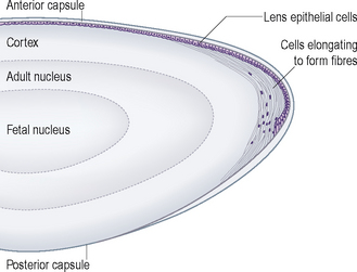
The anterior capsule of the lens is in contact with the posterior iris, which glides over it as the pupil size alters. The posterior capsule has attachments to the vitreous (as the hyaloideocapsular ligament). The adult lens consists of primary and secondary lens fibres. The primary ones differentiate from the posterior lens epithelium at an early stage of eye development and this differentiation is induced by the embryological retina. These primary fibres elongate and fill the lens vesicle to form a solid sphere. In the adult these primary fibres form the central ‘nucleus’ of the lens. Abnormalities of this early differentiation will manifest as nuclear cataracts. The posterior lens epithelium disappears as the lens matures, leaving just a very thin posterior lens capsule. The anterior epithelial cells are located beneath the anterior lens capsule and actively divide throughout life. As they multiply, they move towards the lens periphery and when they reach the equatorial region they start to elongate to form the secondary lens fibres, one arm of which stretches into the anterior cortex and the other into the posterior cortex (Figure 39.2). New secondary fibres are constantly produced throughout life, resulting in progressive compression of the lens nucleus. The anterior lens capsule is produced by the anterior epithelium throughout life and gets thicker as the animal ages. The secondary lens fibres are partially responsible for the normal ageing change of senile nuclear sclerosis. Sclerosis of the lens occurs in the older animal and is not as obvious in the cat as in the dog. It should be differentiated from a true cataract.
Stay updated, free articles. Join our Telegram channel

Full access? Get Clinical Tree


